

In-vivo skeletal muscle mitochondrial function in Klinefelter syndrome
- PMID: 34493629
- PMCID: PMC8712372
- DOI: 10.1136/jim-2021-001966
In-vivo skeletal muscle mitochondrial function in Klinefelter syndrome
Abstract
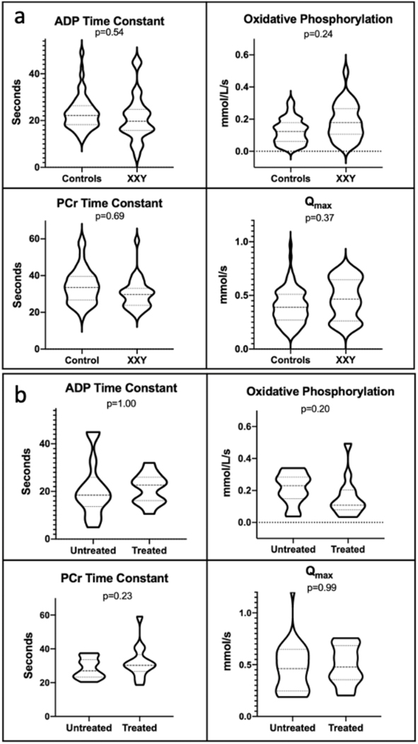
Klinefelter syndrome (XXY) occurs in 1 in 600 males, resulting in testosterone deficiency and a high prevalence of insulin resistance. Testosterone deficiency in men is a known cause of insulin resistance, and mitochondrial dysfunction is hypothesized to mediate this relationship. The aim of this cross-sectional study was to evaluate muscle mitochondrial function in XXY compared with male controls. Twenty-seven boys with XXY (age 14.7±1.8 years) were compared with 87 controls (age 16.9±0.9). In-vivo calf muscle mitochondrial function was assessed via phosphorus magnetic resonance spectroscopy (31P-MRS) following 90 s of isometric 70% maximal exercise. Multiple linear regression was used to compare 31P-MRS outcomes (ADP and phosphocreatine (PCr) time constants, rate of oxidative phosphorylation (Oxphos), and Qmax or the maximal mitochondrial function relative to mitochondrial density) between groups after adjusting for age differences. There were no statistically significant differences in the mitochondrial outcomes of ADP, Oxphos, PCr, and Qmax between the groups. There were also no differences in a sensitivity analysis within the XXY group by testosterone treatment status. In this study, in-vivo postexercise skeletal muscle mitochondrial function does not appear to be impaired in adolescents with XXY compared with controls and is not significantly different by testosterone treatment status in XXY.
Keywords: muscle; skeletal; testosterone.
© American Federation for Medical Research 2022. No commercial re-use. See rights and permissions. Published by BMJ.
Conflict of interest statement
Competing interests: None declared.
Figures
References
-
- Granato S, Barbaro G, Di Giorgio MR, et al. Epicardial fat: the role of testosterone and lipid metabolism in a cohort of patients with Klinefelter syndrome. Metabolism 2019;95:21–6. - PubMed
-
- Accardo G, Amoresano Paglionico V, Di Fraia R, et al. Management of cardiovascular complications in Klinefelter syndrome patients. Expert Rev Endocrinol Metab 2019;14:145–52. - PubMed
-
- Lizarazo AH, McLoughlin M, Vogiatzi MG. Endocrine aspects of Klinefelter syndrome. Curr Opin Endocrinol Diabetes Obes 2019;26:60–5. - PubMed
Publication types
MeSH terms
Substances
Grants and funding
LinkOut - more resources
Full Text Sources
Medical
