

Π-GISANS: probing lateral structures with a fan shaped beam
- PMID: 34493764
- PMCID: PMC8423805
- DOI: 10.1038/s41598-021-97112-x
Π-GISANS: probing lateral structures with a fan shaped beam
Abstract
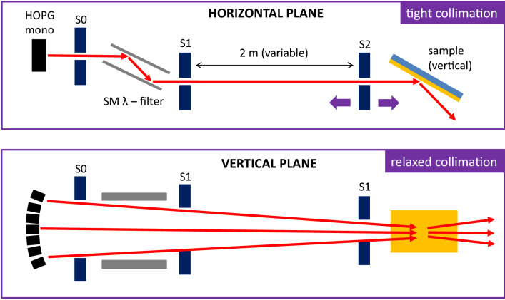
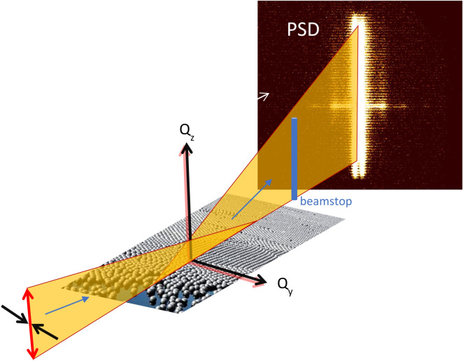
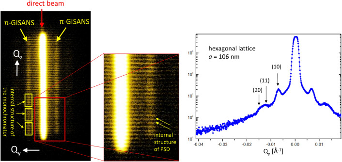
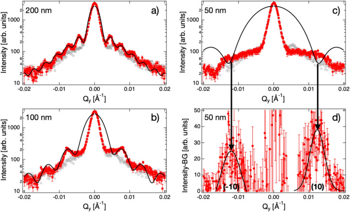
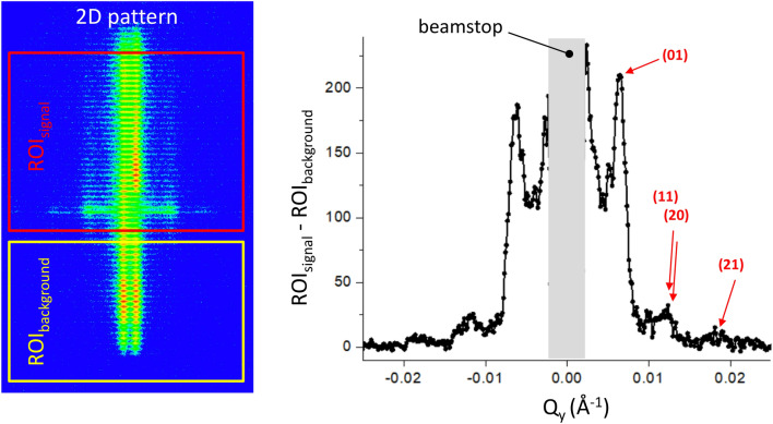
We have performed grazing incidence neutron small angle scattering using a fan shaped incident beam focused along one dimension. This allows significantly reduced counting times for measurements of lateral correlations parallel to an interface or in a thin film where limited depth resolution is required. We resolve the structure factor of iron inclusions in aluminium oxide and show that the ordering of silica particles deposited on a silicon substrate depends on their size. We report hexagonal packing for 50 nm but not for 200 nm silica spheres deposited by a modified Langmuir-Schaefer method on a silicon substrate. For the 200 nm particles we extract the particles shape from the form factor. Moreover, we report dense packing of the particles spread on a free water surface. We name this method π-GISANS to highlight that it differs from GISANS as it gives lateral information while averaging the in-depth structure.
© 2021. The Author(s).
Conflict of interest statement
The authors declare no competing interests.
Figures
References
-
- Giessibl FJ. Advances in atomic force microscopy. Rev. Mod. Phys. 2003;75:949. doi: 10.1103/RevModPhys.75.949. - DOI
-
- Egerton RF, editor. Physical Principles of Electron Microscopy. New York: Springer; 2016.
-
- Hüfner S, editor. Photoelectron Spectroscopy. New York: Springer; 2003.
-
- Levine JR, Cohen JB, Chung YW, Georgopoulos P. J. Appl. Cryst. 1989;22:528–532. doi: 10.1107/S002188988900717X. - DOI
-
- Hexemer A, Bras W, Glossinger J, Schaible E, Gann E, Kirian R, MacDowell A, Church M, Rude B, Padmore H. J. Phys. Conf. Ser. 2010;247:012007. doi: 10.1088/1742-6596/247/1/012007. - DOI
Grants and funding
LinkOut - more resources
Full Text Sources
Research Materials
Miscellaneous
