Review
. 2022 Feb;126(2):165-173.
doi: 10.1038/s41416-021-01543-2. Epub 2021 Sep 7.

Lymphoid-specific helicase in epigenetics, DNA repair and cancer

Affiliations
Review

Lymphoid-specific helicase in epigenetics, DNA repair and cancer

Xiangyu Chen et al. Br J Cancer. 2022 Feb.

Abstract

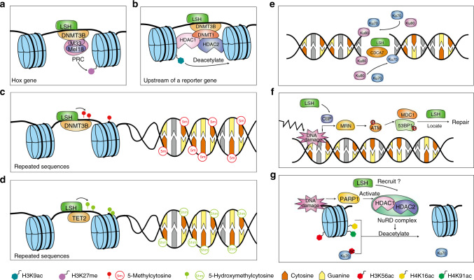
Lymphoid-specific helicase (LSH) is a member of the SNF2 helicase family of chromatin-remodelling proteins. Dysfunctions or mutations in LSH causes an autosomal recessive disease known as immunodeficiency-centromeric instability-facial anomaly (ICF) syndrome. Interestingly, LSH participates in various aspects of epigenetic regulation, including nucleosome remodelling, DNA methylation, histone modifications and heterochromatin formation. Further, LSH plays a crucial role during DNA-damage repair, specifically during double-strand break (DSB) repair, since murine LSH was shown to be essential for non-homologous end joining (NHEJ) and homologous recombination (HR). Accordingly, overexpression of LSH drives tumorigenesis and malignancy. On the other hand, LSH homologs stabilise the genome. Thus, LSH might be implemented as a biomarker for various cancer types and potential target molecule to develop therapeutic strategies against them. In this review, we focus on the role of LSH in orchestrating chromatin rearrangements, such as DNA methylation and histone modifications, as well as in DNA-damage repair. Changes in chromatin structure may facilitate gene expression signatures that cause malignant transformation. We summarise recent findings of LSH in cancers and raise critical open questions for further studies.


Conflict of interest statement

The authors declare no competing interests. This manuscript has been read and approved by all the authors, and has not been submitted to or is not under consideration for publication elsewhere.

Figures

Fig. 1
Fig. 1. LSH is a key regulator of chromatin structure and DNA-damage repair.
a LSH interacts with DNMT3B and PRC components to promote H3K27 methylation of Hox genes and therefore silences their expression during cell development. b LSH recruits DNMT3B, DNMT1, HDAC1 and HDAC2 to deacetylate H3K9ac, acting as a histone transcriptional repressor. c LSH participates in the methylation of repetitive sequences, which are essential components of heterochromatin. LSH helps DNMT3B locate to DNA and methylate centromeric repeats, transposons and gene promoters rich with CpG sites. d LSH upregulates TET2 and forms a complex with it to maintain the genomic 5-hmC level. In response to DNA-damage, mammalian cells initiate DDR and downstream pathways. For DSB, there are two major pathways to repair the damage, NHEJ and HR. Besides, two minor pathways, rd-NHEJ and MMEJ, can ligate ends. As a member of the SWI/SNF chromatin remodeler family, LSH helps to remodel chromatin and facilitates repairing molecules that binds to damaged sites. e LSH and CDCA7 participate in C-NHEJ, in which they recruit protein Ku70 and Ku80 to the damaged sites for afterwards repairing. f In HR, after DNA damage, MRN and ATM are activated, impacting various substrates. LSH recruits MDC1 and 53BP1 protein to targeted sites, facilitating the repairing process. g In MMEJ, HDAC1 and HDAC2 participate in DNA repair by deacetylating H3K56, H4K16 and H4K91, as well as Ku70. Considering that LSH cooperates with HDAC1 and HDAC2 in epigenetic modulation of histone, there exists a possibility that LSH recruits HDAC1 and HDAC2 in response to DNA damage.
Fig. 2
Fig. 2. The expression of LSH in various cancers.
Data analysed in this figure were extracted from the TCGA database (https://portal.gdc.cancer.gov/). The overall survival rate graph of NSCLC is from the study conducted by Mao et al., and the usage of the graph was approved (PMID: 30094095) [95]. a LSH is overexpressed in many types of cancers. Here displays mRNA level in several kinds of cancers with the statistical difference (P < 0.05) between cancerous samples and normal ones. Information on LSH mRNA expression is from TCGA. To conduct the overall analysis, we filtered data with restriction, including transcriptome profiling of data category, gene expression qualification of data type, RNA-seq of experimental strategy and HTSeq-FPKM of workflow type. The nine presented graphs and the related projects are (bladder cancer: TCGA-BLCA), (breast cancer: TCGA-BRCA), (oesophageal carcinoma: TCGA-ESCA), (kidney cancer: TCGA-KIRC, TCGA-KIRP and TCGA-KICH), (liver cancer: TCGA-LIHC), (lung cancer: TCGA-LUAD and TCGA-LUSC), (thyroid carcinoma: TCGA-THCA) and (uterine corpus endometrial carcinoma: TCGA-UCEC). The gene expression levels were computed in an unpaired t-test using GraphPad Prism (v8.0.2). b The survival time correlates with the expression level of LSH. There is a correlation between patients’ survival time and the mRNA level of LSH. The elevated level of LSH mRNA could be a risk factor in breast cancer, oesophageal carcinoma, NSCLC and ovarian cancer. In contrast, high level of LSH mRNA might be a protective factor in head-neck squamous cell carcinoma, liver hepatocellular carcinoma, pancreatic ductal adenocarcinoma and pheochromocytoma and paraganglioma. Data were respectively extracted: breast cancer from TCGA-BRCA, oesophageal carcinoma from TCGA-ESCA, NSCLC from the study conducted by Mao et al. [95], ovarian cancer from TCGA-OV, head-neck squamous cell carcinoma from TCGA-HNSC, liver hepatocellular carcinoma from TCGA- LIHC, pancreatic adenocarcinoma from TCGA-PAAD, and pheochromocytoma and paraganglioma from TCGA-PCPG. Overall survival Kaplan–Meier estimate was completed based on public TCGA data with GraphPad Prism (v8.0.2) and default settings. The outcomes demonstrate that the correlation can be positive or negative, depending on types of cancers. Only curves with P < 0.05 are displayed.
Fig. 3
Fig. 3. The roles of LSH in carcinogenesis.
LSH is elevated in many cancers, which results from the increase of oncogenic factors and the interference with its ubiquitination. AhR binds to the Lsh gene promotor, and SP1 assembles at the upstream of the TSS, elevating the levels of LSH. Besides, there is a positive correlation between FOXM1 and LSH mRNAs. The lncRNA GIAT4RA impedes LSH binding with UCHL3, therefore hinders the ubiquitination of LSH. Increased LSH impacts metabolism, progress and even the fate of cancerous cells. LSH upregulates GLUTs and FADSs, activates TP53 protein and curbs FH. As a consequence, it promotes the Warburg effect, engages in TP53-related lipid catabolism, promotes TP53-related proline catabolism, and suppresses TCA, which motivates IKKα to switch binding sites and consequently, to promote EMT. Increased levels of LSH increase the levels of oncogenic proteins, including GINS4, E2F3 and CENPF, preserving the aggressive phenotype. Also, it inhibits ferroptosis by increasing the levels of the lncRNA LINC00336 and decreasing P53RRA mRNA. The lncRNA LINC00336 stabilises CBS, enhancing cell resistance to ferroptosis. The decline of P53RRA mRNA downregulates Fe2+ and ROS, and as a result, inhibits ferroptosis. However, the relationship between TP53 and LSH needs a cleared elucidation since P53RRA mRNA upregulates TP53 in an upstream manner. In summary, the anomalous expression of LSH accelerates carcinogenesis and leads to worse clinical outcomes.
Fig. 4
Fig. 4. Domains of LSH homologs.
Five homologs from animals, plants and fungi are depicted above. Protein names and species are listed at the left. Conserved domains are labelled. SNF-rel refers to SNF2 related domain; Helicase_C refers to Helicase conserved C-terminal domain.

References

    1. Cavalli G, Heard E. Advances in epigenetics link genetics to the environment and disease. Nature. 2019;571:489–99. - PubMed
    1. Boland M, Nazor K, Loring J. Epigenetic regulation of pluripotency and differentiation. Circulation Res. 2014;115:311–24. - PMC - PubMed
    1. Dobersch, S, Rubio, K, Barreto, G. Pioneer factors and architectural proteins mediating embryonic expression signatures in cancer. Trends Mol Med. 2019. 10.1016/j.molmed.2019.01.008. - PubMed
    1. Singh AK, Mueller-Planitz F. Nucleosome positioning and spacing: from mechanism to function. J Mol Biol. 2021;433:166847. - PubMed
    1. Flaus A, Martin D, Barton G, Owen-Hughes T. Identification of multiple distinct Snf2 subfamilies with conserved structural motifs. Nucleic Acids Res. 2006;34:2887–905. - PMC - PubMed

Publication types

LinkOut - more resources