

Forgotten rhythms? Revisiting the first evidence for rhythms in cognition
- PMID: 34494328
- PMCID: PMC9542866
- DOI: 10.1111/ejn.15450
Forgotten rhythms? Revisiting the first evidence for rhythms in cognition
Abstract
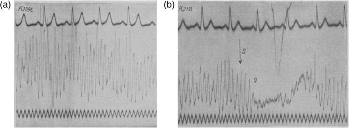
Practically every neuroscientist knows that human brain rhythms were first recorded in the 1920s by Hans Berger, who coined the term 'alpha waves' for the regular activity of around 10 cycles per second that was clearly visible in many of his recordings. Almost 100 years later, alpha rhythms are still the subject of active investigation and continue to intrigue researchers. What we have perhaps forgotten though, is the clever experimentation that was carried out during the first decades of electroencephalogram (EEG) research, often using sophisticated, custom-made analysis and stimulation devices. Here, I review selected findings from the early EEG literature regarding the character, origin, and meaning of human brain rhythms, beginning with Berger's publications and then focusing on the use of regular visual stimulation as a tool to understand intrinsic brain rhythms. It is clear that many of these findings are still relevant to open questions about the role of rhythmic brain activity. In addition, they also contain some general lessons for contemporary neuroscientists, meaning that there is great value in looking back at these forgotten publications.
Keywords: Hans Berger; alpha rhythms; good scientific practice; history of neuroscience; photic driving.
© 2021 The Author. European Journal of Neuroscience published by Federation of European Neuroscience Societies and John Wiley & Sons Ltd.
Conflict of interest statement
The author declares no conflict of interest.
Figures
References
-
- Berger, H. (1929). Über das Elektroenkephalogramm des Menschen. Archiv für Psychiatrie und Nervenkrankheiten, 87(1), 527–570. 10.1007/BF01797193 - DOI
-
- Berger, H. (1930). Über das Elektrenkephalogramm des Menschen. II. Journal für Psychologie und Neurologie, 40, 160–179. [no doi, not available online?]
-
- Berger, H. (1931). Über das Elektrenkephalogramm des Menschen. Dritte Mitteilung. Archiv für Psychiatrie und Nervenkrankheiten, 94, 16–60. 10.1007/BF01835097 - DOI
-
- Berger, H. (1932). Über das Elektrenkephalogramm des Menschen. Vierte Mitteilung. Archiv für Psychiatrie und Nervenkrankheiten, 97, 6–26. 10.1007/BF01815532 - DOI
Publication types
MeSH terms
LinkOut - more resources
Full Text Sources
