

Impact of radiofrequency ablation (RFA) on bone quality in a murine model of bone metastases
- PMID: 34495961
- PMCID: PMC8425524
- DOI: 10.1371/journal.pone.0256076
Impact of radiofrequency ablation (RFA) on bone quality in a murine model of bone metastases
Abstract
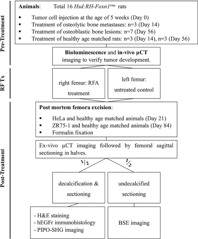
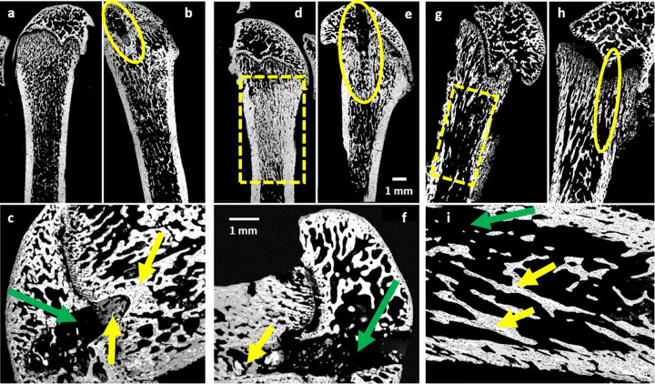
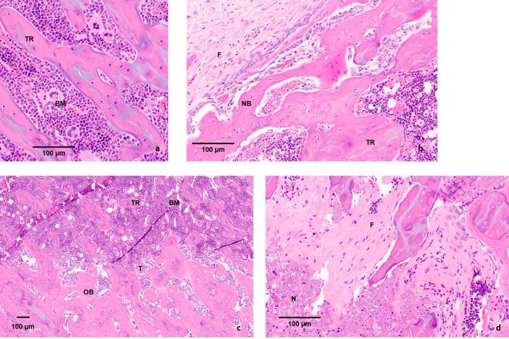
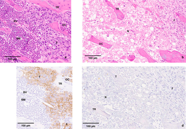
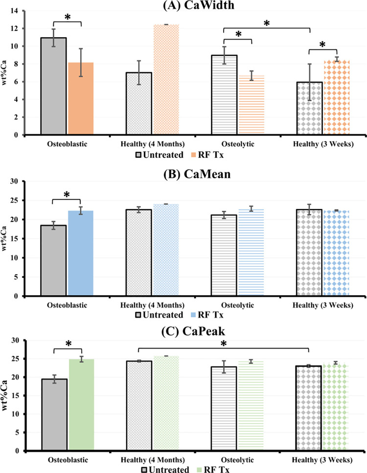
Thermal therapies such as radiofrequency ablation (RFA) are gaining widespread clinical adoption in the local treatment of skeletal metastases. RFA has been shown to successfully destroy tumor cells, yet the impact of RFA on the quality of the surrounding bone has not been well characterized. RFA treatment was performed on femora of rats with bone metastases (osteolytic and osteoblastic) and healthy age matched rats. Histopathology, second harmonic generation imaging and backscatter electron imaging were used to characterize changes in the structure, organic and mineral components of the bone after RFA. RFA treatment was shown to be effective in targeting tumor cells and promoting subsequent new bone formation without impacting the surrounding bone negatively. Mineralization profiles of metastatic models were significantly improved post-RFA treatment with respect to mineral content and homogeneity, suggesting a positive impact of RFA treatment on the quality of cancer involved bone. Evaluating the impact of RFA on bone quality is important in directing the growth of this minimally invasive therapeutic approach with respect to fracture risk assessment, patient selection, and multimodal treatment planning.
Conflict of interest statement
The authors have declared that no competing interests exist.
Figures
Similar articles
-
Radiofrequency ablation of experimental bone metastases in nude rats.Anticancer Res. 2008 Mar-Apr;28(2A):879-85. Anticancer Res. 2008. PMID: 18507032
-
Radiofrequency Ablation Provides Rapid and Durable Pain Relief for the Palliative Treatment of Lytic Bone Metastases Independent of Radiation Therapy: Final Results from the OsteoCool Tumor Ablation Post-Market Study.Cardiovasc Intervent Radiol. 2023 May;46(5):600-609. doi: 10.1007/s00270-023-03417-x. Epub 2023 Apr 3. Cardiovasc Intervent Radiol. 2023. PMID: 37012392 Free PMC article.
-
Phase I/II Study of Radiofrequency Ablation for Painful Bone Metastases: Japan Interventional Radiology in Oncology Study Group 0208.Cardiovasc Intervent Radiol. 2018 Jul;41(7):1043-1048. doi: 10.1007/s00270-018-1944-x. Epub 2018 Apr 12. Cardiovasc Intervent Radiol. 2018. PMID: 29675772 Clinical Trial.
-
Radiofrequency thermoablation (RFA) and radiotherapy (RT) combined treatment for bone metastases: a systematic review.Eur Rev Med Pharmacol Sci. 2021 May;25(10):3647-3654. doi: 10.26355/eurrev_202105_25930. Eur Rev Med Pharmacol Sci. 2021. PMID: 34109573
-
The use of radiofrequency ablation in the treatment of musculoskeletal tumors.J Am Acad Orthop Surg. 2009 Dec;17(12):737-43. doi: 10.5435/00124635-200912000-00002. J Am Acad Orthop Surg. 2009. PMID: 19948698 Review.
Cited by
-
Progression of Femoral Osteolytic Metastases after Intramedullary Nailing and Subsequent Salvage Techniques.Cancers (Basel). 2024 Aug 10;16(16):2812. doi: 10.3390/cancers16162812. Cancers (Basel). 2024. PMID: 39199585 Free PMC article.
-
Mechanical bone strength decreases considerably after microwave ablation-Ex-vivo and in-vivo analysis in sheep long bones.PLoS One. 2023 Oct 12;18(10):e0292177. doi: 10.1371/journal.pone.0292177. eCollection 2023. PLoS One. 2023. PMID: 37824490 Free PMC article.
-
Recent Advances in Minimally Invasive Management of Osteolytic Periacetabular Skeletal Metastases.Semin Intervent Radiol. 2024 Jul 10;41(2):154-169. doi: 10.1055/s-0044-1787165. eCollection 2024 Apr. Semin Intervent Radiol. 2024. PMID: 38993598 Free PMC article. Review.
-
Bone Mass Changes Following Percutaneous Radiofrequency Ablation, Osteoplasty, Reinforcement, and Internal Fixation of Periacetabular Osteolytic Metastases.J Clin Med. 2023 Jul 11;12(14):4613. doi: 10.3390/jcm12144613. J Clin Med. 2023. PMID: 37510728 Free PMC article.
-
Comparison of Percutaneous Interventional Ablation-Osteoplasty-Reinforcement-Internal Fixation (AORIF), Long Intramedullary Nailing, and Hemiarthroplasty for the Treatment of Focal Metastatic Osteolytic Lesions in the Femoral Head and Neck.Cardiovasc Intervent Radiol. 2023 May;46(5):649-657. doi: 10.1007/s00270-023-03425-x. Epub 2023 Apr 13. Cardiovasc Intervent Radiol. 2023. PMID: 37052716
References
-
- Wong DA, Fornasier VL, MacNab I. Spinal metastases: the obvious, the occult, and the impostors. Spine (Phila Pa 1976). 1990;15(1):1–4. Epub 1990/01/01. . - PubMed
Publication types
MeSH terms
LinkOut - more resources
Full Text Sources
Medical
