. 2021 Sep 8;15(9):e0009736.
doi: 10.1371/journal.pntd.0009736. eCollection 2021 Sep.

Leptospira interrogans biofilm formation in Rattus norvegicus (Norway rats) natural reservoirs

Affiliations

Leptospira interrogans biofilm formation in Rattus norvegicus (Norway rats) natural reservoirs

Ana Amélia Nunes Santos et al. PLoS Negl Trop Dis. .

Abstract

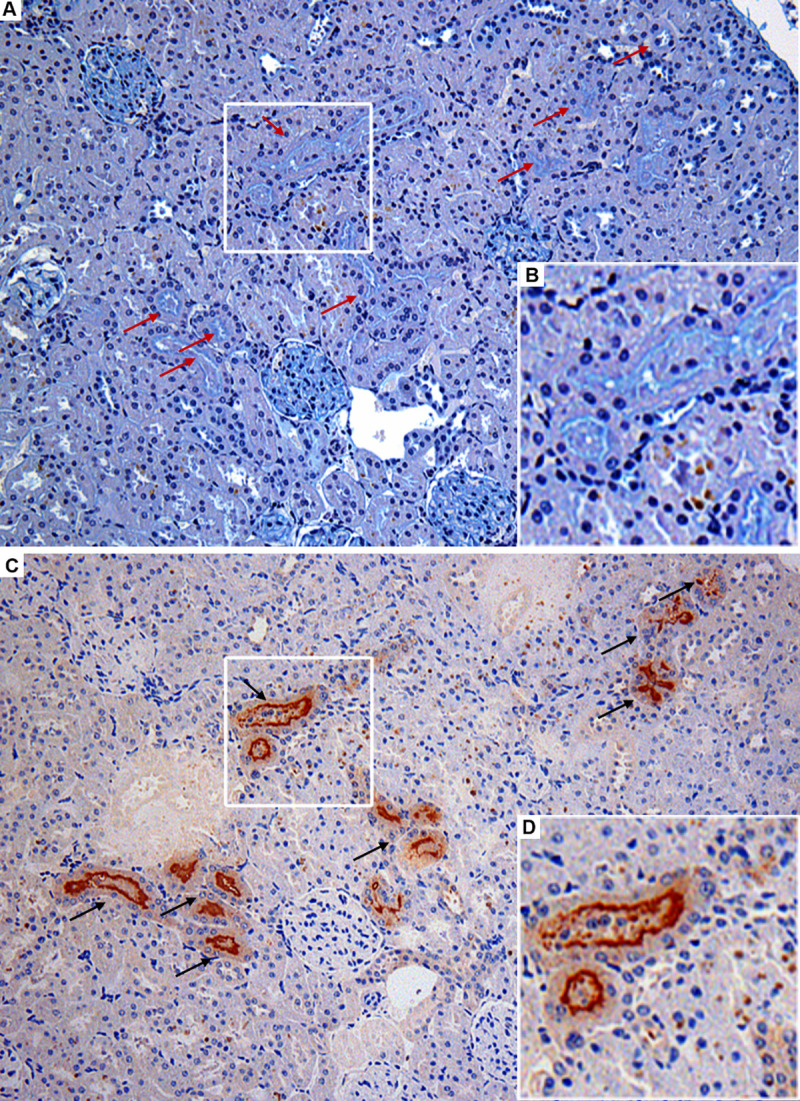
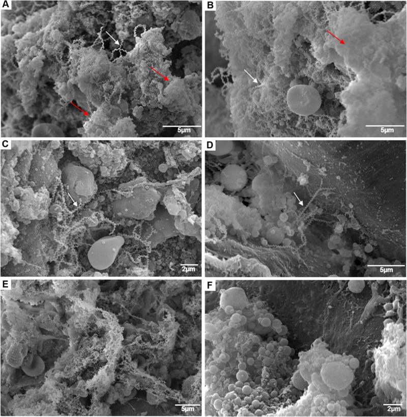
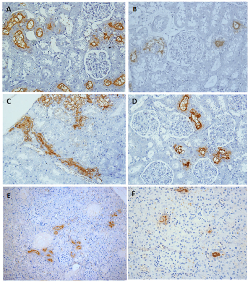
Rattus norvegicus (Norway rat) is the main reservoir host of pathogenic Leptospira, the causative agent of leptospirosis, in urban environments. Pathogenic Leptospira forms biofilms in the environment, possibly contributing for bacterial survival and maintenance. Nonetheless, biofilms have not yet been studied in natural animal reservoirs presenting leptospiral renal carriage. Here, we described biofilm formation by pathogenic Leptospira inside the renal tubules of R. norvegicus naturally infected and captured in an urban slum endemic for leptospirosis. From the 65 rats carrying Leptospira in their kidneys, 24 (37%) presented biofilms inside the renal tubules. The intensity of leptospiral colonization in the renal tubules (OR: 1.00; 95% CI 1.05-1.1) and the type of occlusion pattern of the colonized renal tubules (OR: 3.46; 95% CI 1.20-9.98) were independently associated with the presence of Leptospira biofilm. Our data showed that Leptospira interrogans produce biofilms during renal chronic colonization in rat reservoirs, suggesting a possible role for leptospiral biofilms in the pathogenesis of leptospirosis and bacterial carriage in host reservoirs.


Conflict of interest statement

The authors have declared that no competing interests exist.

Figures

Fig 1
Fig 1. Histopathological investigation of leptospiral infection and biofilm formation in kidney serial sections of wild Rattus norvegicus naturally infected with pathogenic Leptospira interrogans.
(A) Alcian Blue (AB) positively stained renal tubules (red arrows) observed in light turquoise blue, indicating the presence of biofilm matrix; insert B with detail of biofilm staining. (C) Renal tubules positive for IHC anti-L. interrogans demonstrating leptospiral colonization of proximal tubules (black arrows); insert D with detail of colonized tubule. Note that the serial sections showed in A and D are from the same region of one rat kidney, evidencing the co-localization of tubules concomitantly positive in AB (red arrows) and IHC (black arrows). Magnification, x 200.
Fig 2
Fig 2. Scanning electron microscopy (SEM) of leptospiral renal biofilm and its matrix in wild naturally colonized Rattus norvegicus.
A and B–SEM of colonized kidney with ruthenium red (RR) showed Leptospira agglomerates (white arrows) surrounded by an anionic exopolysaccharidic matrix (red arrows) inside the renal tubules. C and D–SEM of colonized kidney without RR, where leptospires are evidenced agglomerated (C) or isolated (D), without the presence of the matrix. E–SEM using RR of R. norvegicus negative control. F–SEM without RR of R. norvegicus negative control.
Fig 3
Fig 3. Patterns of kidney colonization marking of naturally infected Rattus norvegicus.
Immunohistochemical representative images of kidney with (A) high intensity of colonized tubules (CTs); (B) low intensity of CTs; (C) partial to complete pattern of CT lumen occlusion; (D) marking restricted to the membrane of renal epithelium (partial occlusion); (E) agglomerated CTs distributed in the renal cortex; (F) isolated CTs distributed in the cortex. A, B, C and D: magnification, x 400. E and F: magnification, x 200.

References

    1. Ko AI, Goarant C, Picardeau M. Leptospira: the dawn of the molecular genetics era for an emerging zoonotic pathogen. Nat Rev Microbiol [Internet]. 2009;7(10):736–47. Available from: doi: 10.1038/nrmicro2208 - DOI - PMC - PubMed
    1. Costa F, Hagan JE, Calcagno J, Kane M, Torgerson P, Martinez-Silveira MS, et al.. Global Morbidity and Mortality of Leptospirosis: A Systematic Review. PLoS Negl Trop Dis. 2015;9(9):0–1. doi: 10.1371/journal.pntd.0003898 - DOI - PMC - PubMed
    1. Casanovas-Massana A, Costa F, Riediger IN, Cunha M, de Oliveira D, Mota DC, et al.. Spatial and temporal dynamics of pathogenic Leptospira in surface waters from the urban slum environment. Water Res [Internet]. 2018;130:176–84. Available from: doi: 10.1016/j.watres.2017.11.068 - DOI - PMC - PubMed
    1. Reis RB, Ribeiro GS, Felzemburgh RDM, Santana FS, Mohr S, Melendez AXTO, et al.. Impact of environment and social gradient on Leptospira infection in urban slums. PLoS Negl Trop Dis. 2008;2(4):11–8. doi: 10.1371/journal.pntd.0000228 - DOI - PMC - PubMed
    1. Casanovas-Massana A, Hamond C, Santos LA, de Oliveira D, Hacker KP, Balassiano I, et al.. Leptospira yasudae sp. Nov. and Leptospira stimsonii sp. nov., two new species of the pathogenic group isolated from environmental sources. Int J Syst Evol Microbiol. 2020;70(3):1450–6. doi: 10.1099/ijsem.0.003480 - DOI - PMC - PubMed

Publication types