. 2021 Sep;1(9):e235.
doi: 10.1002/cpz1.235.

Development of Keratinocyte Cell Lines Containing Extrachromosomal Human Papillomavirus Genomes

Affiliations

Development of Keratinocyte Cell Lines Containing Extrachromosomal Human Papillomavirus Genomes

Tami L Coursey et al. Curr Protoc. 2021 Sep.

Abstract

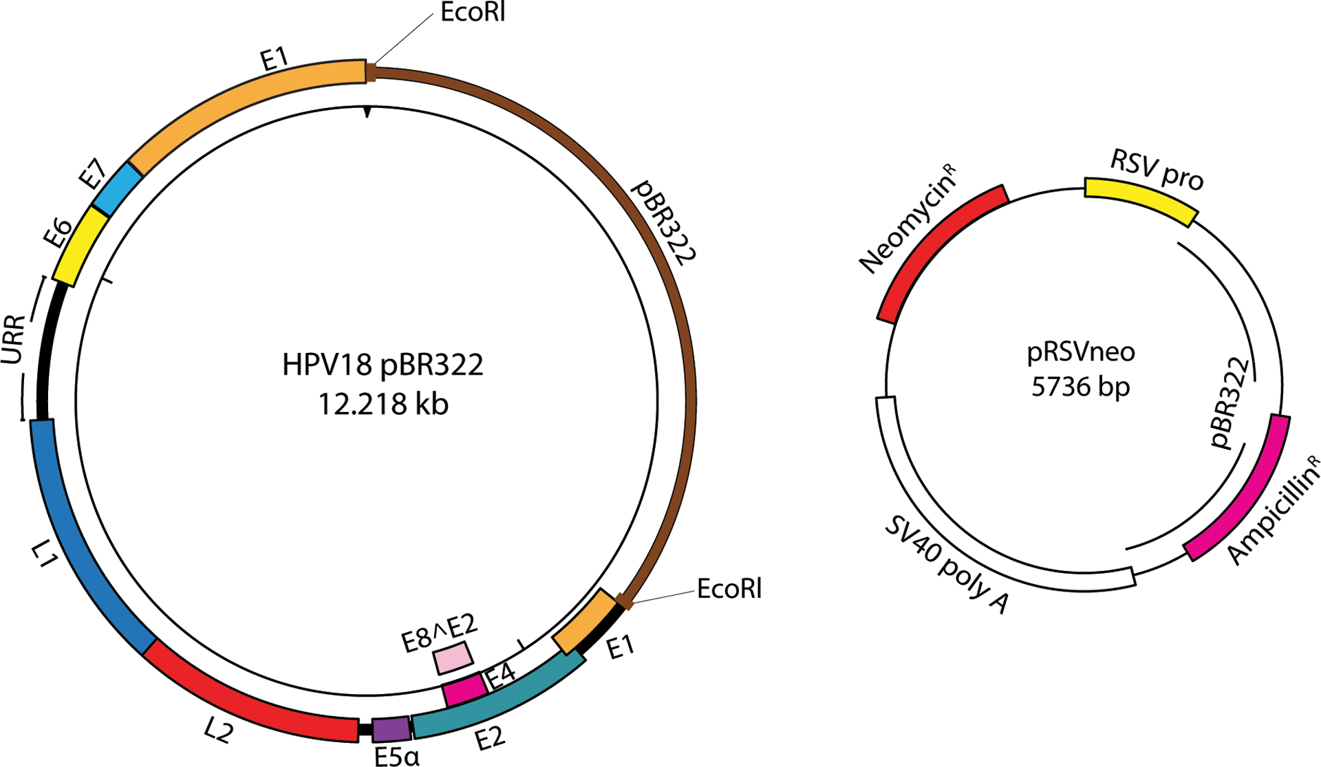
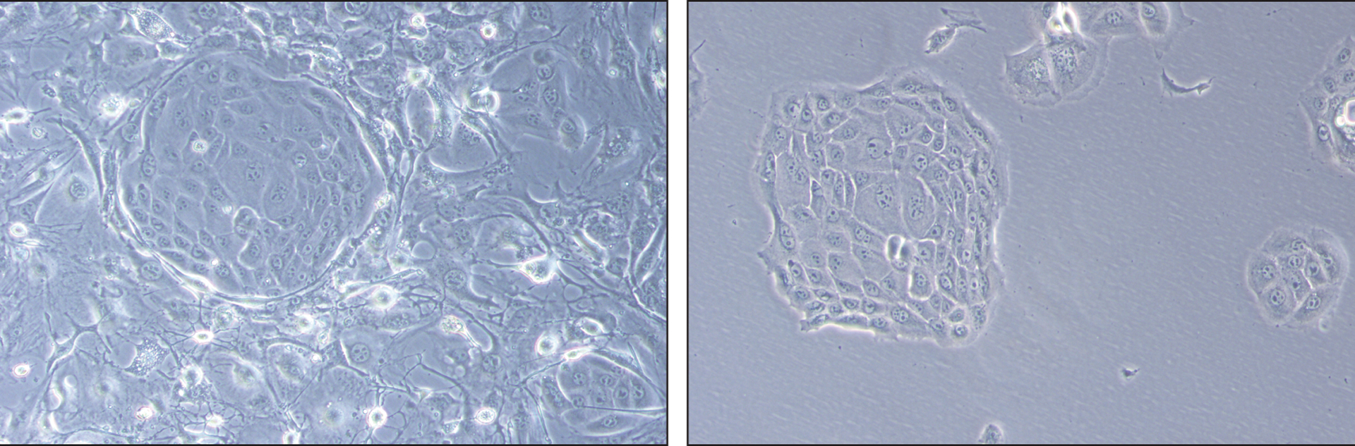
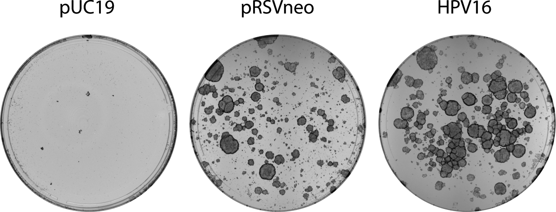
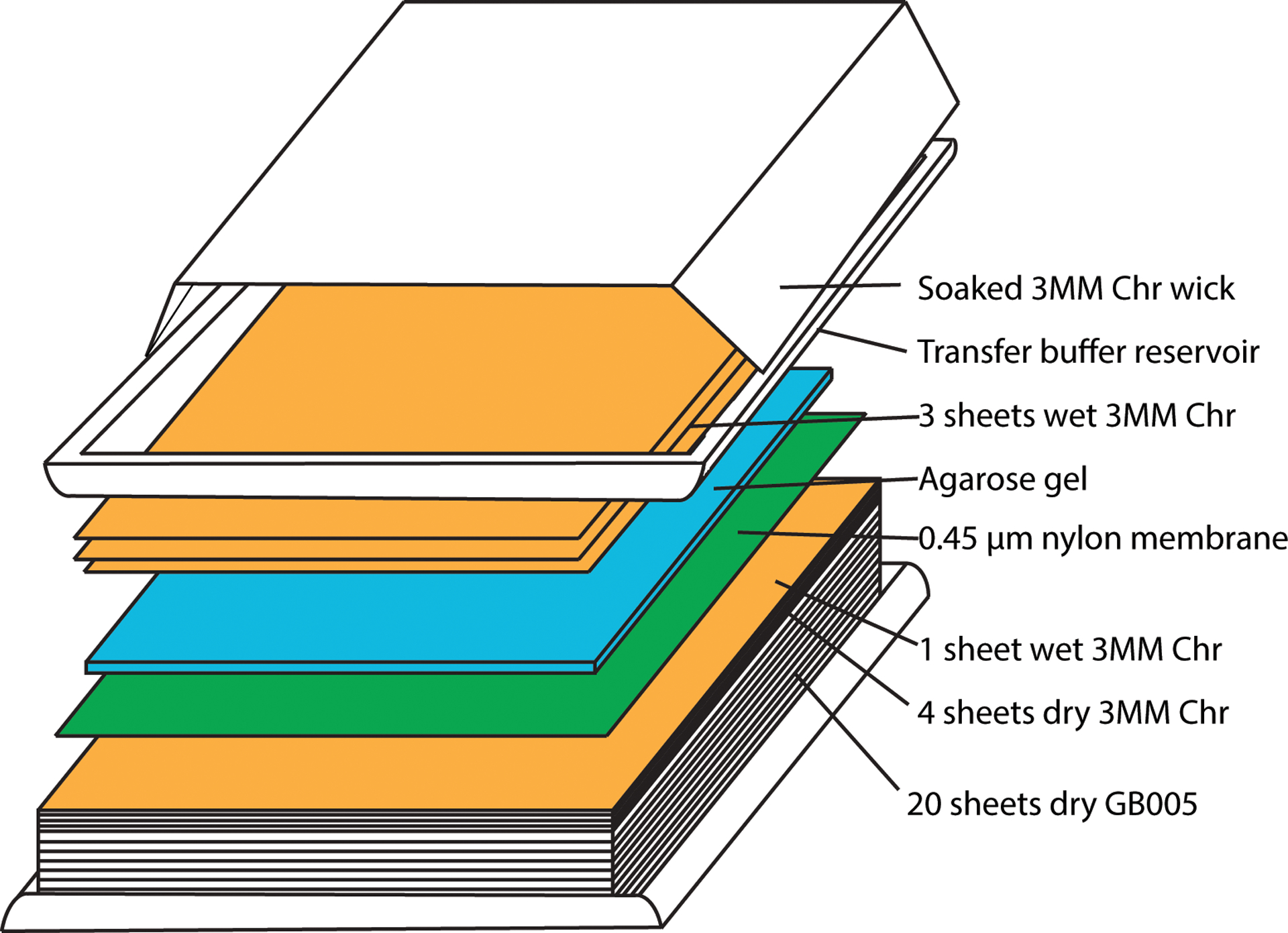
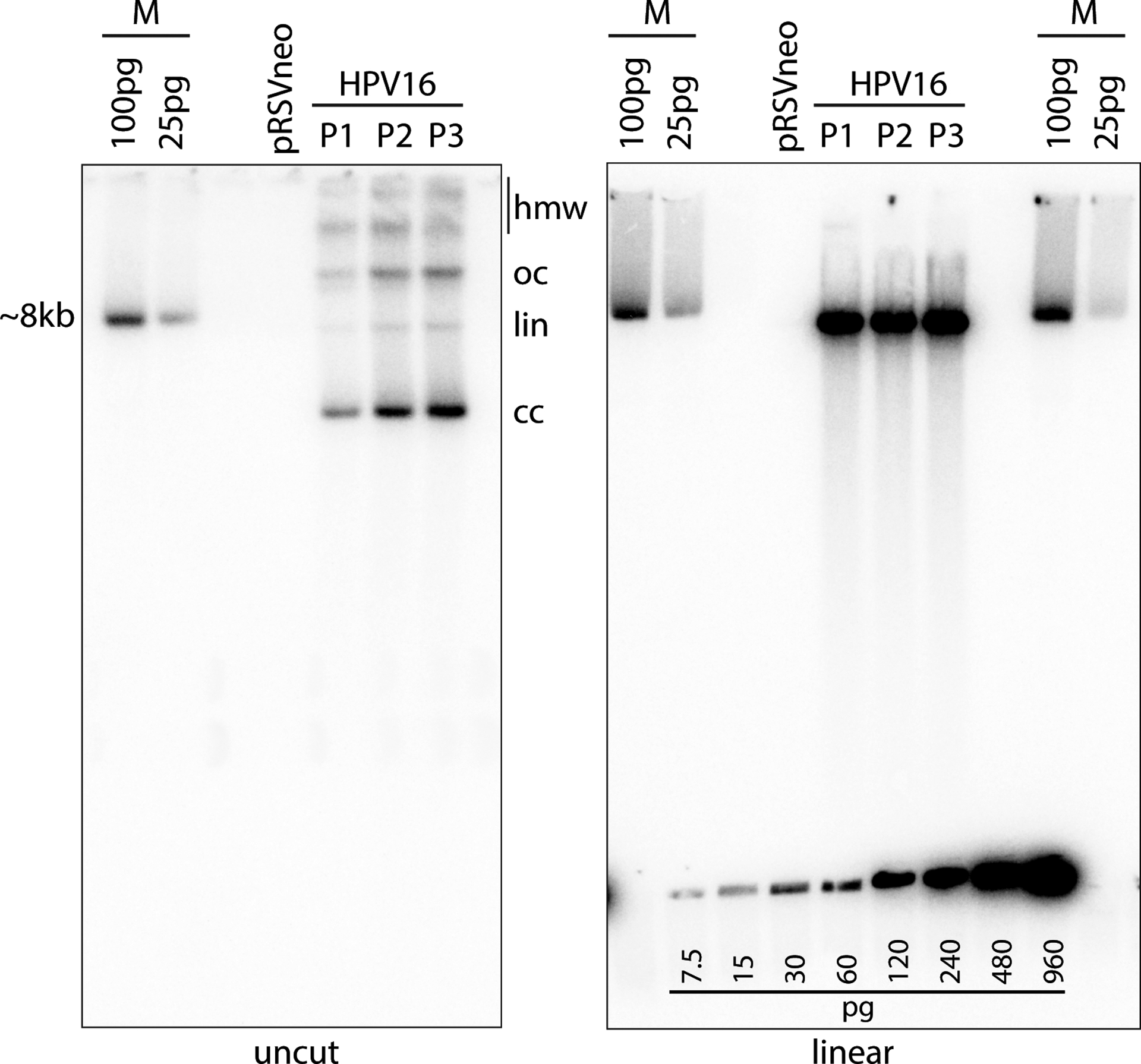
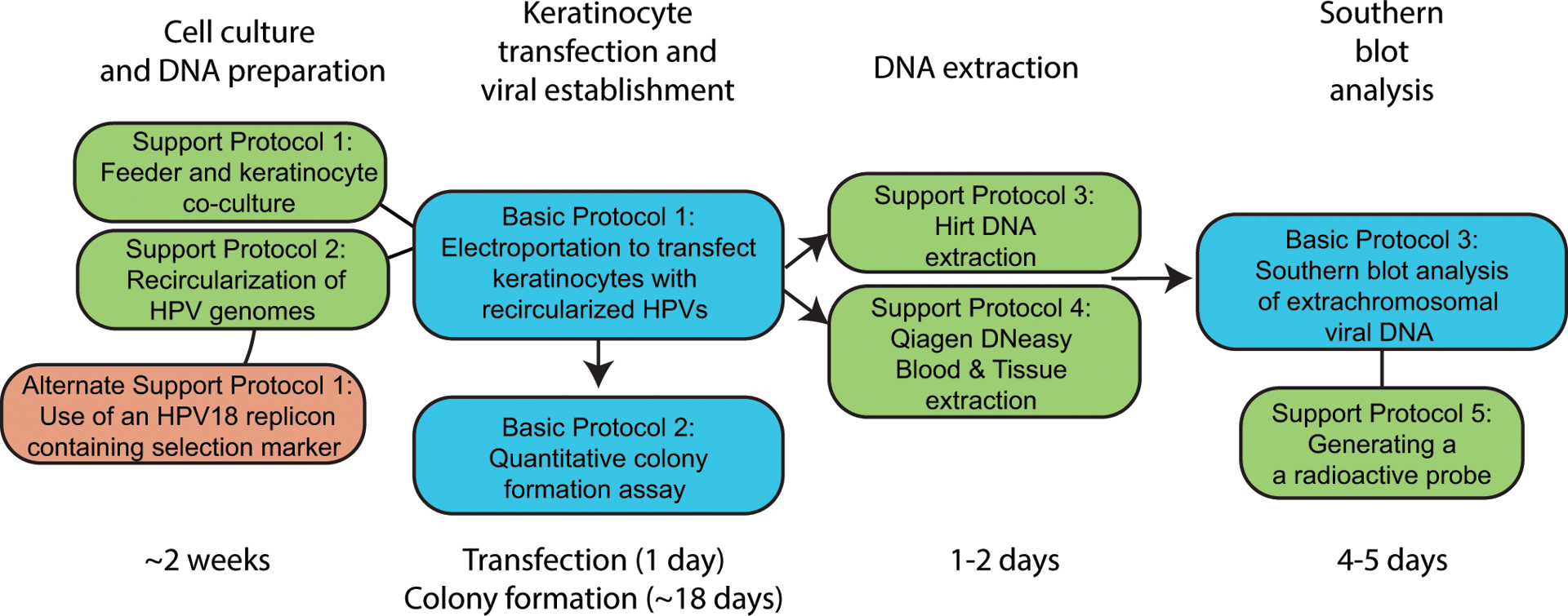
Human papillomaviruses (HPVs) cause persistent infections in stratified cutaneous and mucosal epithelia. In these infections, the viral DNA replicates as low-copy-number, extrachromosomal, double-stranded-DNA circular plasmids in the nucleus of the dividing basal cells. When the infected cells begin the process of differentiation, the viral DNA amplifies to a high copy number and virions are assembled in the superficial cells. To study HPV DNA replication, our laboratory generates primary keratinocyte cell lines that contain replicating extrachromosomal HPV genomes. Here, we describe protocols to culture human keratinocytes, to transfect viral DNA into cells using electroporation, to determine the efficiency of genome establishment in cells with a colony-forming assay, and to measure the copy number and extrachromosomal status of viral genomes using Southern blotting. These methods can be used to study DNA replication of different oncogenic Alphapapillomavirus HPV types. Published 2021. This article is a U.S. Government work and is in the public domain in the USA. Basic Protocol 1: Electroporation to transfect keratinocytes with recircularized HPV genomes Alternate Protocol: Use of HPV replicon containing selection marker in keratinocyte transfection Support Protocol 1: Rheinwald-Green method of co-culture of irradiated J2 3T3 feeders and human keratinocytes Support Protocol 2: Recircularization of HPV genomes Basic Protocol 2: Quantitative colony formation assay to measure the efficiency of HPV genome establishment Basic Protocol 3: Southern blot analysis of extrachromosomal viral DNA Support Protocol 3: Hirt extraction of low-molecular-weight DNA Support Protocol 4: Qiagen DNeasy Blood & Tissue DNA extraction Support Protocol 5: Generation of a 32 P-labeled HPV DNA probe.

Keywords: HPV; Southern blot; extrachromosomal DNA; human papillomavirus; keratinocyte; replication.


Figures

Figure 1.
Figure 1.
Plasmid maps of HPV18 (cloned in pBR322) and selectable marker plasmid (pRSVneo). The HPV genome is cloned in pBR322, a bacterial plasmid vector, for propagation in bacteria. A restriction digest (the EcoRl site is highlighted in red to release the HPV18 genome from pBR322 vector) releases the HPV genome from this vector for HPV genome recircularization (Support Protocol 2) prior to transfection (Basic Protocol 1). The pRSVneo plasmid is co-transfected with the recircularized HPV genome and enables transient G418 selection of transfected cells (Basic Protocol 1).
Figure 2.
Figure 2.
Keratinocyte colonies co-cultured with fibroblast feeders. An image was taken of a colony of proliferative keratinocytes pushing surrounding feeders away as the colony continues to grow. Feeders and keratinocyte cells are indicated. The same colony is visualized on the right after fibroblast feeders are removed with Versene.
Figure 3.
Figure 3.
HPV keratinocyte colonies stained with methylene blue 18 days after electroporation. HFK colonies resulting from the method described in Basic Protocol 1 were stained with methylene blue as described in Basic Protocol 2 and imaged on a Syngene white box imager. Comparing the number of colonies formed by transfecting with HPV16 and pRSVneo versus pRSVneo and pUC19, can help determine whether the concentration of G418 used in selection should be increased or decreased. A plate of control plasmid(1.g.pUC19) transfected keratinocytes can help determine the effectiveness of the G418 drug used.
Figure 4.
Figure 4.
Assembly of capillary transfer unit for southern blot analysis.
Figure 5.
Figure 5.
Southern blot analysis. In this example, DNA was extracted from cells passed from the colonies shown in Figure 2 using the Qiagen DNeasy Blood and Tissue Kit (Support Protocol 4). Three micrograms of total cellular DNA was digested with Hindlll (cleaves cellular DNA but does not cut HPV16 viral genome) or BamHl (cuts the viral genome once) and analyzed by Southern blot analysis as detailed in Basic Protocol 3. The left gel shows the HPV DNA isomers detected by the radioactive probe (see Supporting Protocol 3): supercoiled, covalently closed (cc), linear (lin) DNA, open circular/nicked (oc) DNA, and high molecular weight (hmw) forms that may include concatemers. Linearized viral DNA digested with BamHl runs as a single ~8kb band as shown in the gel on the right. The cloned linear HPV16 genome, cleaved from the bacterial vector, is used as a size marker (M). A DNA standard was also created using the HPV16 size marker and was loaded along the bottom of the gel on the right. This DNA standard can be used to estimate the viral DNA copy number. The calculated loaded DNA standard amount should be adjusted to account for the cloned bacterial plasmid and compared to the viral DNA signal in the cell DNA lane. The amount of DNA in a single human cell is ~6pg. Therefore, 3μg cell DNA represents 5 × 10⁵ cells. Each pg of viral DNA (7900bp dsDNA) contains 1.155 ×10⁵ molecules. Thus, the number of viral DNA copies per cell can be easily calculated.
Figure 6.
Figure 6.
Timeline and overview of protocols. Basic protocols are highlighted in blue, support protocols in green, and alternative protocols in red. General timelines for protocols are listed below.

References

    1. Allen-Hoffmann BL, Schlosser SJ, Ivarie CA, Sattler CA, Meisner LF, & O’Connor SL (2000). Normal growth and differentiation in a spontaneously immortalized near-diploid human keratinocyte cell line, NIKS. J Invest Dermatol, 114(3), 444–455. doi:10.1046/j.1523-1747.2000.00869.x - DOI - PubMed
    1. Boshart M, Gissmann L, Ikenberg H, Kleinheinz A, Scheurlen W, & zur Hausen H (1984). A new type of papillomavirus DNA, its presence in genital cancer biopsies and in cell lines derived from cervical cancer. EMBO J, 3(5), 1151–1157. Retrieved from https://www.ncbi.nlm.nih.gov/pubmed/6329740 - PMC - PubMed
    1. Chapman S, Liu X, Meyers C, Schlegel R, & McBride AA (2010). Human keratinocytes are efficiently immortalized by a Rho kinase inhibitor. J Clin Invest, 120(7), 2619–2626. doi:10.1172/JCI42297 - DOI - PMC - PubMed
    1. Coursey TL, Van Doorslaer K, & McBride AA (2021). Regulation of HPV18 Genome Replication, Establishment and Persistence by Sequences in the Viral Upstream Regulatory Region. J Virol, JVI0068621. doi:10.1128/JVI.00686-21 - DOI - PMC - PubMed
    1. Fehrmann F, & Laimins LA (2005). Human papillomavirus type 31 life cycle: methods for study using tissue culture models. Methods Mol Biol, 292, 317–330. doi:10.1385/1-59259-848-x:317 - DOI - PubMed

LinkOut - more resources