. 2021 Sep 9;385(11):996-1004.
doi: 10.1056/NEJMoa2100440.

Somatic PIK3CA Mutations in Sporadic Cerebral Cavernous Malformations

Affiliations

Somatic PIK3CA Mutations in Sporadic Cerebral Cavernous Malformations

Matthieu Peyre et al. N Engl J Med. .

Abstract

Background: Cerebral cavernous malformations (CCMs) are common sporadic and inherited vascular malformations of the central nervous system. Although familial CCMs are linked to loss-of-function mutations in KRIT1 (CCM1), CCM2, or PDCD10 (CCM3), the genetic cause of sporadic CCMs, representing 80% of cases, remains incompletely understood.

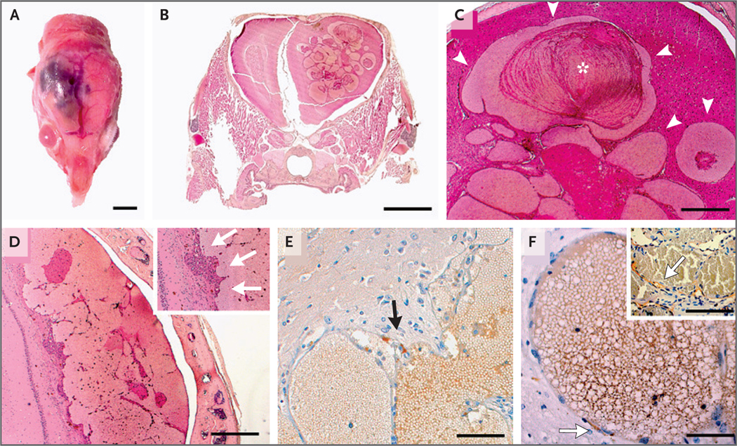
Methods: We developed two mouse models harboring mutations identified in human meningiomas with the use of the prostaglandin D2 synthase (PGDS) promoter. We performed targeted DNA sequencing of surgically resected CCMs from patients and confirmed our findings by droplet digital polymerase-chain-reaction analysis.

Results: We found that in mice expressing one of two common genetic drivers of meningioma - Pik3ca H1047R or AKT1 E17K - in PGDS-positive cells, a spectrum of typical CCMs develops (in 22% and 11% of the mice, respectively) instead of meningiomas, which prompted us to analyze tissue samples from sporadic CCMs from 88 patients. We detected somatic activating PIK3CA and AKT1 mutations in 39% and 1%, respectively, of lesion tissue from the patients. Only 10% of lesions harbored mutations in the CCM genes. We analyzed lesions induced by the activating mutations Pik3ca H1074R and AKT1 E17K in mice and identified the PGDS-expressing pericyte as the probable cell of origin.

Conclusions: In tissue samples from sporadic CCMs, mutations in PIK3CA were represented to a greater extent than mutations in any other gene. The contribution of somatic mutations in the genes that cause familial CCMs was comparatively small. (Funded by the Fondation ARC pour la Recherche contre le Cancer and others.).


Figures

Figure 1
Figure 1. Characterization of Mouse Pik3caH1047R CCMs.
Panel A is a whole-mount view of a dissected brain showing a cerebral cavernous malformation (CCM) in the brain stem of a 1-month-old PGDSCre;Pik3caH1047R mouse (scale bar, 1 mm). Panel B shows a section through the lesion in which blood-filled caverns are visible (scale bar, 200 μm). Panel C shows in situ hybridization, with prostaglandin D2 synthase (Pgds) messenger RNA expression in pericavernomatous cells (scale bar, 50 μm). Panels D, E, and F show representative examples of CCMs located in the brain stem of 3.2-month-old (Panel D), 1-month-old (Panel E), and 2.8-month-old (Panel F) PGDSCre;Pik3caH1047R mice (scale bar, 2 mm). Panel G shows dilated capillaries in normal brain parenchyma (asterisks) (scale bar, 100 μm). Panel H shows the formation of noncoalescent intraparenchymal telangiectasias with preserved spherical structure (asterisks) (scale bar, 100 μm). Panel I shows fusion of adjacent telangiectasias with a thin band of residual brain parenchyma between the two caverns (arrowheads) (scale bar, 100 μm). Panel J shows immunohistochemical detection of PGDS-positive cells around dilated blood vessels (scale bar, 50 μm). The inset shows an example of a PGDS-positive cell bordering a cavern (arrow). Panel K shows Masson’s trichrome staining of a brain-stem lesion with thin borders separating the caverns (scale bar, 2 mm). The inset shows an example of a more mature human cavernoma, with thick blue collagen fibers separating the caverns. Panel L shows an example of intraluminal thrombi found in some CCMs (scale bar, 100 μm).
Figure 2.
Figure 2.. Characterization of Mouse AKT1E17K CCMs.
Panel A shows the macroscopic appearance of a right cerebral cortical lesion in a 13-month-old PGDStv-a;RCAS-AKT1E17K mouse (scale bar, 5 mm). Panel B shows an example of a frontal lobe CCM in a 13-month-old PGDStv-a;RCAS-AKT1E17K mouse (scale bar, 2 mm). Panel C shows the lesions, which consist of tortuous dilated vascular channels lined with a single layer of endothelium without intervening parenchyma (arrowheads), similar to human CCMs. Cavities are filled with erythrocytes along with intraluminal and parietal thrombi (asterisk); also evident are clusters of siderophages, lymphocyte infiltrates, and reactive gliosis, indicating past hemorrhages (scale bar, 500 μm). Panel D shows a cerebral cortical lesion formed by the coalescence of several small cavities with intraluminal thrombi (scale bar, 500 μm). The inset shows endothelial cells of neovessels (arrows) that line the cavities. Panel E shows FLAG immunohistochemical staining, which detected AKT1E17K expression in pericavernous cells of a mouse CCM, in the arachnoid cells of meningothelial proliferations, and in cells bordering CCMs (arrow), suggesting that the lesions are associated with AKT1E17K expression (scale bar, 100 μm). Panel F shows PGDS immunohistochemical staining, which indicates the presence of scattered PGDS-positive cells (arrow) boarding the intraparenchymal caverns without any contact with the meninges (scale bar, 50 μm). The inset shows a human CCM lesion with PGDS-positive cells bordering vascular channels (arrow).
Figure 3
Figure 3. Detection of Mutations in Human Sporadic CCMs.
Panel A shows the allele frequencies of variants in PIK3CA, CCM1, CCM2, CCM3, and AKT1, determined on the basis of either the percentage of sequence reads that contained variants on next-generation sequencing (NGS) or the fractional abundance of variants on digital droplet polymerase-chain-reaction (ddPCR) analysis, in human sporadic CCM samples. Panel B shows clinical characteristics, including sample type (fresh-frozen or formalin-fixed, paraffin-embedded [FFPE] tissue), the sex of the patient, CCM location, and the specific activating mutation detected. For some samples, insufficient tissue remained for ddPCR analysis. Panel C shows examples of PIK3CA- and AKT1-mutant human CCMs. Axial three-dimensional T1-weighted MRI of the insular cavernoma harboring PIK3CA, CCM1, and AKT1 mutations (Patient 5) is shown, followed by axial MRIs of the PIK3CA-mutant CCMs with the highest fractional abundance of mutant reads. Patient numbers are shown beneath the scans.

Comment in

  • Cellular Origin of Sporadic CCMs.
    Ren J, Hong T, Zhang H. Ren J, et al. N Engl J Med. 2022 Mar 31;386(13):1290-1291. doi: 10.1056/NEJMc2117812. N Engl J Med. 2022. PMID: 35353971 No abstract available.
  • Cellular Origin of Sporadic CCMs.
    Betsholtz C, Gaengel K, Mäkinen T. Betsholtz C, et al. N Engl J Med. 2022 Mar 31;386(13):1291. doi: 10.1056/NEJMc2117812. N Engl J Med. 2022. PMID: 35353972 No abstract available.

References

    1. Awad IA, Polster SP. Cavernous angiomas: deconstructing a neurosurgical disease. J Neurosurg 2019; 131: 1–13. - PMC - PubMed
    1. Rigamonti D, Hadley MN, Drayer BP, et al. Cerebral cavernous malformations: incidence and familial occurrence. N Engl J Med 1988; 319: 343–7. - PubMed
    1. Riant F, Bergametti F, Ayrignac X, Boulday G, Tournier-Lasserve E. Recent insights into cerebral cavernous malformations: the molecular genetics of CCM. FEBS J 2010; 277: 1070–5. - PubMed
    1. D’Angelo R, Marini V, Rinaldi C, et al. Mutation analysis of CCM1, CCM2 and CCM3 genes in a cohort of Italian patients with cerebral cavernous malformation. Brain Pathol 2011; 21:215–24. - PMC - PubMed
    1. D’Angelo R, Alafaci C, Scimone C, et al. Sporadic cerebral cavernous malformations: report of further mutations of CCM genes in 40 Italian patients. Biomed Res Int 2013; 2013: 459253. - PMC - PubMed

Publication types