. 2021 Sep 28;60(38):2888-2901.
doi: 10.1021/acs.biochem.1c00365. Epub 2021 Sep 8.

Significant Loop Motions in the SsoPTP Protein Tyrosine Phosphatase Allow for Dual General Acid Functionality

Affiliations

Significant Loop Motions in the SsoPTP Protein Tyrosine Phosphatase Allow for Dual General Acid Functionality

Justin Pinkston et al. Biochemistry. .

Abstract

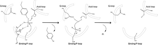
Conformational dynamics are important factors in the function of enzymes, including protein tyrosine phosphatases (PTPs). Crystal structures of PTPs first revealed the motion of a protein loop bearing a conserved catalytic aspartic acid, and subsequent nuclear magnetic resonance and computational analyses have shown the presence of motions, involved in catalysis and allostery, within and beyond the active site. The tyrosine phosphatase from the thermophilic and acidophilic Sulfolobus solfataricus (SsoPTP) displays motions of its acid loop together with dynamics of its phosphoryl-binding P-loop and the Q-loop, the first instance of such motions in a PTP. All three loops share the same exchange rate, implying their motions are coupled. Further evidence of conformational flexibility comes from mutagenesis, kinetics, and isotope effect data showing that E40 can function as an alternate general acid to protonate the leaving group when the conserved acid, D69, is mutated to asparagine. SsoPTP is not the first PTP to exhibit an alternate general acid (after VHZ and TkPTP), but E40 does not correspond to the sequence or structural location of the alternate general acids in those precedents. A high-resolution X-ray structure with the transition state analogue vanadate clarifies the role of the active site arginine R102, which varied in structures of substrates bound to a catalytically inactive mutant. The coordinated motions of all three functional loops in SsoPTP, together with the function of an alternate general acid, suggest that catalytically competent conformations are present in solution that have not yet been observed in crystal structures.


Figures

Figure 1.
Figure 1.
Chemical steps of the PTP-catalyzed reaction. The WPD-loop assumes a catalytically active closed conformation with the general acid (Asp) in position to protonate the leaving group during formation of the phosphoenzyme intermediate. In the second step this intermediate is hydrolyzed. After the phosphate product is released, the open WPD-loop conformation becomes favored.
Figure 2.
Figure 2.
Alignment of the catalytic regions of PTP1B (red), YopH (blue), and SsoPTP (green) each with bound vanadate, showing the common geometries of their active sites. The Q-loop is characterized by a conserved glutamine, shown in sticks. This residue positions a nucleophilic water molecule in the second catalytic step as shown in Figure 1. The acid-loop in PTPs contains a conserved general acid, shown in sticks, (D69 in SsoPTP) which protonates the leaving group in the first catalytic step. The binding-loop, also referred to as the P-loop, contains the nucleophilic cysteine, shown in sticks at the bottom of the figure which attacks the phosphorus in the first step of the mechanism.
Figure 3:
Figure 3:
The substrate p-nitrophenyl phosphate (pNPP) showing the positions at which kinetic isotope effects were measured: the bridge oxygen atom, the position of bond cleavage, (V/K)bridge; the nitrogen atom in leaving group, (V/K).
Figure 4.
Figure 4.
SsoPTP complexed with the transition state analog vanadate. The R102 interactions in this complex are typical of those observed in other PTPs indicative of bridging interactions with two oxygen atoms of the -PO3 (phosphoryl) group. Positions of the conserved general acid D69 and the potential general acid E40 are shown. The pH where this structure and previous ones have been obtained is well above optimal activity, where E40 is ionized and found in a salt bridge with R102 and not in a location amenable to protonate the leaving group. Structural reorganization, potentially facilitated at lower pH where E40 should be largely protonated, would be required to bring it into proximity to fill the role implied by the kinetic data. Alternatively, given the structure with pNPP bound to the inactive mutant (Figure S1), E40 may act in concert with R102 as the proton donor.
Figure 5.
Figure 5.
2D 1H-15N-TROSY spectra of SsoPTP at 35 °C. (Top) ligand-free SsoPTP. Unassigned residues are shown absent a label. (Bottom) SsoPTP in the presence of 200-fold molar excess of tungstate at 35 °C. Residues corresponding to each backbone amide H-N correlation are labeled. Resonances that appear in the presence of tungstate are G98, G101, R102, T103, and G104.
Figure 6.
Figure 6.
The assigned residues are mapped onto the crystal structure of the tungstate (shown in CPK rendering) bound SsoPTP as shown as a solid ribbon, and the unassigned residues are shown in dashed ribbon. Proline residues are colored in black. Secondary structures are colored as follows: α-helices are in red, β-strands are in yellow, and loop regions are represented in green. PDB ID: 2I6M
Figure 7.
Figure 7.
The overlay of 2D 1H-15N-TROSY spectrum of the SsoPTP during the tungstate titration at 35 °C. The ligand-free (free) spectrum is shown in red, and the tungstate bound spectra (enzyme-ligand ratio) are shown in orange (1:0.5), yellow (1:3.6), green (1.21), blue (1:100), and purple (1:200). Residues that appear in the presence of tungstate are located at the active site, and identified by the blue circle. Residues that disappear in the presence of tungstate are also near the active site and are highlighted with the red circle.
Figure 8.
Figure 8.
Conformational motions of ligand-free SsoPTP as probed by R experiments. The dynamic residues, D69 and V72, in the acid loop are shown onto the crystal structure of the ligand-free SsoPTP as red spheres. Relaxation data was collected at 600 MHz. Global fit to the two-site exchange equation (eq 3) is represented with the solid line. PDB code: 2I6I
Figure 9.
Figure 9.
Conformational motions of tungstate bound SsoPTP as probed by CPMG relaxation dispersion experiments. (left) 15N-CPMG relaxation dispersion curves of the dynamic residues in tungstate bound SsoPTP are plotted. Global fit to the fast two-site exchange equation represented with solid line. (right) The dynamic residues in the acid, P-, and Q-loop are mapped onto the crystal structure of the tungstate bound SsoPTP as shown as spheres. PDB ID: 2I6M
Figure 10.
Figure 10.
Conformational motions of ligand-free and tungstate bound SsoPTP are probed by R and CPMG experiments. In ligand-free, D69, V72 of the acid loop mobile with a kex = 3140 s−1. In the tungstate bound form, I67, D69, V72 of the acid loop, G99, R102, and T103 of the conserved PTP motif, Y2, and A133, Q135 of the Q-loop are also flexible with kex reduced and = 760 s−1. The dynamic residues are mapped onto the crystal structure of the ligand-free and tungstate bound SsoPTP, as shown in spheres. PDB code: 2I6I (ligand-free), 2I6M (tungstate bound).
Figure 11.
Figure 11.
pH profiles of SsoPTP WT (blue squares), D69N (red circles), E40Q (green diamonds), and D69N E40Q (black triangles). Data were obtained at 25°C in 200mM succinate. D69N, WT, and E40Q all display bell-shaped trends consistent with operational general acid catalysis. The rate reduction seen in the double mutant D69N/E40Q (black), along with the elimination of the basic limb, indicate abolished general acid catalysis.
Figure 12
Figure 12
(A). VHZ (yellow), Tk-PTP (purple), and SsoPTP (green) in complex with metavanadate, vanadate, and vanadate, respectively. (B) Sequence alignment of VHZ, Tk-PTP, and SsoPTP. Structures were obtained from PDB IDs 4ERC, 5Z5A, and 2I6I, respectively. Highlighting in the first column shows the glutamate residue found to function as an alternative general acid in SsoPTP and the corresponding residues in the other two enzymes. The aspartic acids highlighted in the second column are the conserved general acid residues common to all PTPs and found in the same active site locations. In the third column, the nucleophilic cysteine is highlighted in all three proteins. Residues highlighted in yellow in the 4th column show the glutamates that can act as the alternate general acids in VHZ and Tk-PTP and the glutamine in the corresponding location in SsoPTP.

References

    1. Cozzone AJ, Protein phosphorylation in prokaryotes. Annu Rev Microbiol 1988, 42, 97–125. - PubMed
    1. Mustelin T; Feng GS; Bottini N; Alonso A; Kholod N; Birle D; Merlo J; Huynh H, Protein tyrosine phosphatases. Front Biosci 2002, 7, d85–142. - PubMed
    1. Brandao TA; Johnson SJ; Hengge AC, The molecular details of WPD-loop movement differ in the protein-tyrosine phosphatases YopH and PTP1B. Arch Biochem Biophys 2012, 525 (1), 53–9. - PMC - PubMed
    1. Jackson MD; Denu JM, Molecular reactions of protein phosphatases--insights from structure and chemistry. Chem Rev 2001, 101 (8), 2313–40. - PubMed
    1. Zhang ZY; Wang Y; Wu L; Fauman EB; Stuckey JA; Schubert HL; Saper MA; Dixon JE, The Cys(X)5Arg catalytic motif in phosphoester hydrolysis. Biochemistry 1994, 33 (51), 15266–70. - PubMed

Publication types

MeSH terms

Substances