. 2021 Nov 8;31(21):4800-4809.e9.
doi: 10.1016/j.cub.2021.08.030. Epub 2021 Sep 7.

A supernumerary "B-sex" chromosome drives male sex determination in the Pachón cavefish, Astyanax mexicanus

Affiliations

A supernumerary "B-sex" chromosome drives male sex determination in the Pachón cavefish, Astyanax mexicanus

Boudjema Imarazene et al. Curr Biol. .

Abstract

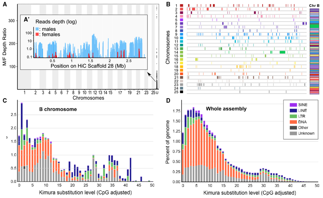
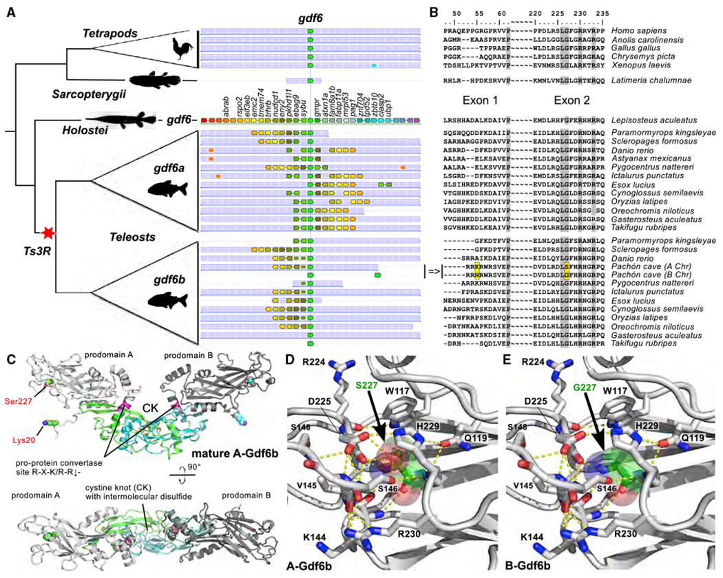
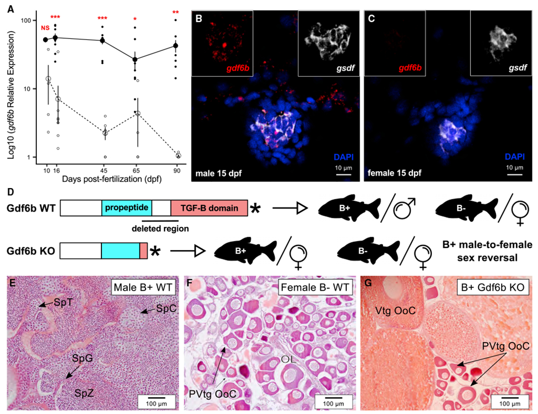
Sex chromosomes are generally derived from a pair of classical type-A chromosomes, and relatively few alternative models have been proposed up to now.1,2 B chromosomes (Bs) are supernumerary and dispensable chromosomes with non-Mendelian inheritance found in many plant and animal species3,4 that have often been considered as selfish genetic elements that behave as genome parasites.5,6 The observation that in some species Bs can be either restricted or predominant in one sex7-14 raised the interesting hypothesis that Bs could play a role in sex determination.15 The characterization of putative B master sex-determining (MSD) genes, however, has not yet been provided to support this hypothesis. Here, in Astyanax mexicanus cavefish originating from Pachón cave, we show that Bs are strongly male predominant. Based on a high-quality genome assembly of a B-carrying male, we characterized the Pachón cavefish B sequence and found that it contains two duplicated loci of the putative MSD gene growth differentiation factor 6b (gdf6b). Supporting its role as an MSD gene, we found that the Pachón cavefish gdf6b gene is expressed specifically in differentiating male gonads, and that its knockout induces male-to-female sex reversal in B-carrying males. This demonstrates that gdf6b is necessary for triggering male sex determination in Pachón cavefish. Altogether these results bring multiple and independent lines of evidence supporting the conclusion that the Pachón cavefish B is a "B-sex" chromosome that contains duplicated copies of the gdf6b gene, which can promote male sex determination in this species.

Keywords: B chromosome; cavefish; gdf6; genome; sex chromosomes; sex determination; sex differentiation, gonads.


Conflict of interest statement

Declaration of interests The authors declare no competing interests

Figures

Figure 1.
Figure 1.. Karyological characterization of male-predominant supernumerary B chromosomes (Bs) in Pachón cave Astyanax mexicanus
(A and B) Representative C-banding patterns of single B+ male (A) and B− female (B) from Pachón cave. The Bs (black arrow) lack C-bands, suggesting that Pachón cavefish Bs are largely euchromatic. See also Figure S1. (C) Boxplots of the average number of Bs per metaphase in Pachón cavefish males and females. Horizontal lines indicate the median, the box indicates the interquartile range (IQR), and the whiskers the range of values that are within 1.5× IQR. Statistical significance between males and females was tested with the Wilcoxon rank test (***p < 0.001). See also Data S1A. (D) Fluorescence in situ hybridization (FISH) of male Pachón cave mitotic metaphase labeled with combined microdissected B probes. Yellow arrows point to the strong labeling of Bs and the small white arrows to the lighter labeling of some different parts of A chromosomes. (E and F) FISH co-labeling of a 1B male (E) and a 1B female (F) metaphase with microdissected B (green) and gdf6b-specific (red) probes. Bs are indicated by yellow arrows and the white arrowheads point to pairs of A chromosome sister chromatids labeled by the gdf6b probe. Only one A-chromosome gdf6b signal was detected in (E). The two gdf6b signals (see inset in E) that were often visible on male metaphases cannot be interpreted as the two different B-gdf6b loci due to their genomic proximity (see Data S1C) and the FISH resolution. (G and H) Synaptonemal complex (SC) analysis showing that Pachón cave Bs (yellow arrow) do not pair with the 25 fully synapsed standard bivalents of A chromosomes. SCs were visualized by anti-SYCP3 antibody (green), the recombination sites were identified by anti-MLH1 antibody (red), and chromosomes were counterstained by DAPI (blue). (G) Merged image. (H) SYCP3 visualization only. Scale bars, 5 μm.
Figure 2.
Figure 2.. Genomic characterization of Pachón cavefish B chromosome (B)
(A) Read depth ratio of male and female Pachón genomic pools showing a strong coverage bias in a single scaffold, Hi_scaffold_28 (enlarged in A’ inset showing male and female read coverage). (B) Karyoplots of the A chromosome regions duplicated on the Pachón cavefish B (Chr B) showing that the Pachón B is made from a complex mosaic of duplicated A chromosomal fragments. (C and D) Comparison of the repeat landscapes of the Pachón B (C) and whole genome including the B (D), showing that the Pachón B has a very different repeat element (color code provided in inset of D) content compared to A chromosomes. Short interspersed repeated sequences, SINEs; long interspersed nuclear elements, LINEs; long terminal repeats, LTRs; DNA repeat elements, DNAs; terminal inverted repeat sequences, TIRs. See also Figures S2A and S2B and Data S1D for additional details.
Figure 3.
Figure 3.. Gdf6b protein evolution and structure
(A) Simplified phylogeny (left panel) and synteny (middle panel) relationships of the gdf6a and gdf6b genes, with the additional B-gdf6b Pachón cavefish duplication, showing that they are duplicated paralogs stemming from the teleost whole-genome duplication (Ts3R). Species names are given on the right side of (B). (B) Corresponding multiple alignments of Gdf6 protein-coding sequences around the A-Gdf6b lysine to B-Gdf6b asparagine switch in Exon 1 (p.Lys60Asn) and the A-Gdf6b serine to B-Gdf6b glycine switch in Exon 2 (p.Ser227Gly). (C) Ribbon plot homology model of A-Gdf6b proprotein dimer (bottom panel view is rotated by 90° around the x axis). The prodomains are shown in light and dark gray, the furin processing site (R274-R-K-R-R278) is indicated in magenta, and the activity-containing mature C-terminal domain is shown in green and cyan. The two residues differing between A-Gdf6b and B-Gdf6b are presented as spheres with their carbon atoms colored in green and cyan. (D and E) Comparative magnifications of the structure of A-Gdf6b (D) and B-Gdf6b (E) around the p.Ser227Gly switch. Amino acid residues interacting with Ser227 or Gly227 are shown as sticks, and hydrogen bonds as yellow stippled lines. As shown, the side chain hydroxyl group of Ser227 (shown with carbon atoms colored in green and transparent spheres highlighting the van der Waals spheres of the atoms) engages in several hydrogen bonds with surrounding residues, e.g., Ser146 and Asp225, thereby stabilizing the tertiary and secondary structure in this region. Upon exchange of Ser227 with a glycine as in B-Gdf6b, these hydrogen bonds are lost, thereby potentially destabilizing this region in the prodomain.
Figure 4.
Figure 4.. Gene expression and functional evidence supporting a role of gdf6b as a potential master sex-determining gene in Pachón cavefish
(A) Expression profiles of gdf6b in male and female trunks during early development from 10 to 90 days post-fertilization (dpf; males, solid line; females, dashed line) showing a significant overexpression in males compared to females starting from 16 dpf. Results are presented as log10 mean ± standard errors; black and white dots represent the individual values of relative expression in males and females, respectively. Statistical significance between males and females was tested with the Wilcoxon rank-sum test (Wilcoxon-Mann-Whitney test) and only significant differences are shown (***p < 0.01; **p < 0.01; *p < 0.05). See also Figure S4A. (B and C) Gonadal expression of gdf6b (in red) and the Sertoli and granulosa supporting cell marker gsdf (in white) in male (B) and female (C) differentiating gonads at 15 dpf showing that gdf6b is specifically expressed in male gonads with no strict colocalization with gsdf in male. See Figure S4B for additional stages of development. Nuclei were stained with DAPI (in blue). Scale bar, 10 μm. (D) Schematic representation of the wild-type (WT) and knockout (KO) Gdf6b proteins and the resulting phenotypes of B+ males and B− females. (E–G) Representative gonadal histology of WT males (E), WT females (F), and Gdf6b KO B+ males showing that Gdf6b KO induces male-to-female sex reversal (G) with ovaries containing vitellogenic (Vtg Ooc) and previtellogenic oocytes (PVtg Ooc), like WT ovaries (F), contrasting with the testis in WT males (E). Ol, ovarian lamellae; SpC, spermatocytes; SpG, spermatogonia; SpT, spermatids; SpZ, spermatozoa. Scale bar, 100 μm.

References

    1. Wright AE, Dean R, Zimmer F, and Mank JE (2016). How to make a sex chromosome. Nat. Commun 7, 12087. - PMC - PubMed
    1. Furman BLS, Metzger DCH, Darolti I, Wright AE, Sandkam BA, Almeida P, Shu JJ, and Mank JE (2020). Sex chromosome evolution: so many exceptions to the rules. Genome Biol. Evol 12, 750–763. - PMC - PubMed
    1. D’Ambrosio U, Alonso-Lifante MP, Barros K, Kovařík A, Mas de Xaxars G, and Garcia S (2017). B-chrom: a database on B-chromosomes of plants, animals and fungi. New Phytol. 216, 635–642. - PubMed
    1. Jones N (2017). New species with B chromosomes discovered since 1980. Nucleus 60, 263–281.
    1. Camacho JPM (2005). B chromosomes. In The Evolution of the Genome, Gregory TR, ed. (Academic Press; ), pp. 223–286.

Publication types