Review
. 2021 Sep 7;33(9):1744-1762.
doi: 10.1016/j.cmet.2021.08.006.

Exosomes as mediators of intercellular crosstalk in metabolism

Affiliations
Review

Exosomes as mediators of intercellular crosstalk in metabolism

Roi Isaac et al. Cell Metab. .

Abstract

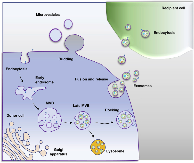
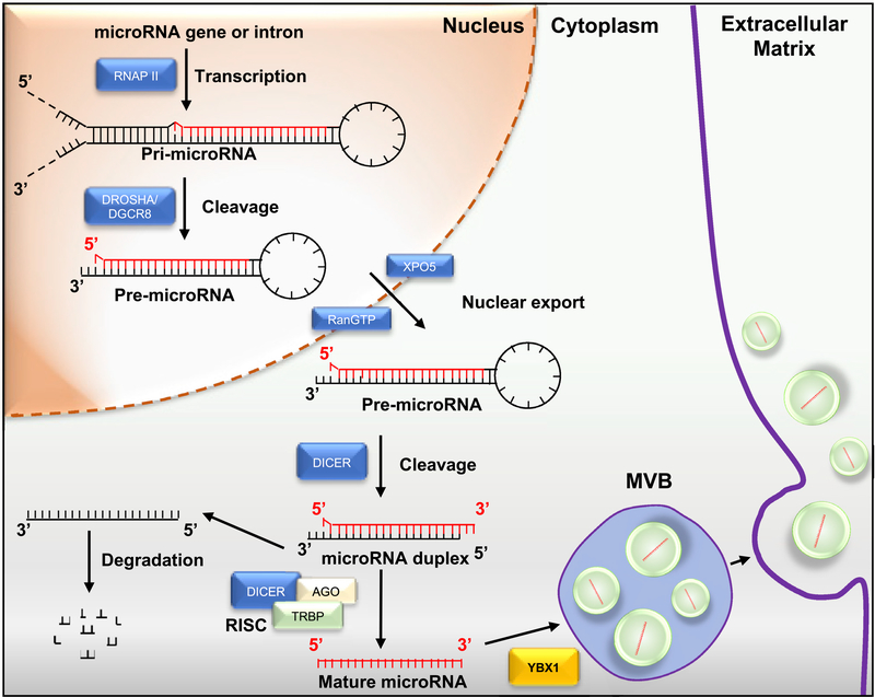
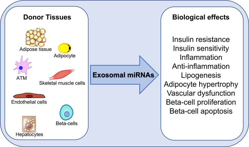
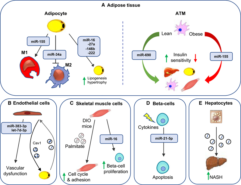
Exosomes are nanoparticles secreted by all cell types and are a large component of the broader class of nanoparticles termed extracellular vesicles (EVs). Once secreted, exosomes gain access to the interstitial space and ultimately the circulation, where they exert local paracrine or distal systemic effects. Because of this, exosomes are important components of an intercellular and intraorgan communication system capable of carrying biologic signals from one cell type or tissue to another. The exosomal cargo consists of proteins, lipids, miRNAs, and other RNA species, and many of the biologic effects of exosomes have been attributed to miRNAs. Exosomal miRNAs have also been used as disease biomarkers. The field of exosome biology and metabolism is rapidly expanding, with new discoveries and reports appearing on a regular basis, and it is possible that potential therapeutic approaches for the use of exosomes or miRNAs in metabolic diseases will be initiated in the near future.


Conflict of interest statement

Declaration of interests The authors declare no competing interests.

Figures

Figure 1.
Figure 1.. Exosome and microvesicle biogenesis pathways
Microvesicles bud directly from the plasma membrane, and exosomes are generated by inward budding of the multivesicular body (MVB) lipid bilayer membrane. MVB fusion with the plasma membrane is a tightly regulated multistep process that includes MVB trafficking along microtubules and docking at the plasma membrane for further exosome release. Alternatively, MVBs can fuse with lysosomes as part of the degradative process. Initially, exosomes bind to the cell surface of recipient cells through protein-protein or receptor-ligand interactions, which can initiate signaling cascades that activate endocytotic pathways. The exosome cargo confers the biologic effects of exosomes on recipient cells.
Figure 2.
Figure 2.. miRNA biogenesis
Pri-miRNAs are hairpin-shaped precursor miRNA molecules transcribed by either RNA polymerase II (RNAPII) from independent genes or introns of protein-coding genes. The pri-miRNAs are cleaved by Drosha/DGCR8 into pre-miRNA and exported to the cytoplasm from the nuclei mediated by Exportin 5 (XPO5) binding. Dicer cleavage generates an miRNA duplex intermediate. Trans-activation-responsive RNA-binding protein (TRBP) and Argonaute (AGO) protein assemble into the RNA-induced silencing complex (RISC). One miRNA strand is transferred to AGO protein, resulting in the formation of RISC. AGO2 binds to one of the miRNA strands to form the mature miRNA, which will be selectively incorporated into exosomes. After miRNA maturation, the RNA-binding protein YBX1 also sorts miRNAs into exosomes.
Figure 3.
Figure 3.. Exosomal miRNA biological effects
Exosomal miRNAs secreted by different metabolic tissues have effects on insulin signaling, inflammation, vascular function, adipocyte biology, and beta cell physiology.
Figure 4.
Figure 4.. Exosomal miRNA effects on recipient cells
Exosomal miRNAs mediate communication between donor cells and recipient cells, playing an important role in metabolic signaling. (A) Adipocyte exosomal miRNAs can modulate macrophage polarization and lipogenesis and hypertrophy in adipocytes. Additionally, adipose tissue macrophage (ATM) exosomes can regulate insulin sensitivity through the delivery of miR-690 to the liver, adipocytes, ATMs, and skeletal muscle. (B) Inflammatory factors induce the release of endothelial cell (EC) exosomes that contain miR-383–3p and let-7d-3p. ECs transfer cav1-containing exosomes to adipocytes, which reciprocate by releasing exosomes taken up by ECs. (C) Skeletal muscle-derived exosomes induce cell cycle and adhesion in skeletal muscle as well as proliferation of isolated mouse beta cells. (D) Cytokine-treated beta cell lines and human islets lead to enhanced secretion of exosomal miR-21–5p. Exosomes from cytokine-treated beta cells induce apoptosis in the recipient cells. (E) Lipotoxic hepatocytes secrete exosomes that activate hepatic stellate cells, promoting the fibrotic NASH phenotype.

References

    1. Alkhouri N, Dixon LJ, and Feldstein AE (2009). Lipotoxicity in nonalcoholic fatty liver disease: not all lipids are created equal. Expert Rev. Gastroenterol. Hepatol 3, 445–451. - PMC - PubMed
    1. Alvarez-Erviti L, Seow Y, Yin H, Betts C, Lakhal S, and Wood MJ (2011). Delivery of siRNA to the mouse brain by systemic injection of targeted exosomes. Nat. Biotechnol 29, 341–345. - PubMed
    1. Amosse J, Durcin M, Malloci M, Vergori L, Fleury A, Gagnadoux F, Dubois S, Simard G, Boursier J, Hue O, et al. (2018). Phenotyping of circulating extracellular vesicles (EVs) in obesity identifies large EVs as functional conveyors of macrophage migration inhibitory factor. Mol. Metab 18, 134–142. - PMC - PubMed
    1. Andreu Z, and Yáñez-Mó M (2014). Tetraspanins in extracellular vesicle formation and function. Front. Immunol 5, 442. - PMC - PubMed
    1. Arraud N, Linares R, Tan S, Gounou C, Pasquet JM, Mornet S, and Brisson AR (2014). Extracellular vesicles from blood plasma: determination of their morphology, size, phenotype and concentration. J. Thromb. Haemost 12, 614–627. - PubMed

Publication types