. 2021 Oct 7;28(10):1726-1739.e6.
doi: 10.1016/j.stem.2021.07.012. Epub 2021 Sep 7.

Antiviral treatment causes a unique mutational signature in cancers of transplantation recipients

Affiliations

Antiviral treatment causes a unique mutational signature in cancers of transplantation recipients

Jurrian K de Kanter et al. Cell Stem Cell. .

Abstract

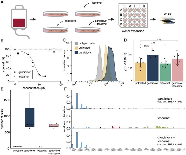
Genetic instability is a major concern for successful application of stem cells in regenerative medicine. However, the mutational consequences of the most applied stem cell therapy in humans, hematopoietic stem cell transplantation (HSCT), remain unknown. Here we characterized the mutation burden of hematopoietic stem and progenitor cells (HSPCs) of human HSCT recipients and their donors using whole-genome sequencing. We demonstrate that the majority of transplanted HSPCs did not display altered mutation accumulation. However, in some HSCT recipients, we identified multiple HSPCs with an increased mutation burden after transplantation. This increase could be attributed to a unique mutational signature caused by the antiviral drug ganciclovir. Using a machine learning approach, we detected this signature in cancer genomes of individuals who received HSCT or solid organ transplantation earlier in life. Antiviral treatment with nucleoside analogs can cause enhanced mutagenicity in transplant recipients, which may ultimately contribute to therapy-related carcinogenesis.

Keywords: antiviral treatment; cancer genomics; cytomegalovirus; ganciclovir; hematopoietic stem cell transplantation; mutational signatures; somatic mutations; therapy-related neoplasms.


Conflict of interest statement

Declaration of interests A.R.H., A.v.L., and R.v.B. are named as inventors on a patent application filed resulting from this work.

Figures

None
Graphical abstract
Figure 1
Figure 1
Mutation accumulation associated with HSCT in humans (A) Schematic of the experimental setup to determine somatic mutations in blood progenitor cells of HSC transplantation (HSCT) donors and recipients. (B) Correlation between the age and the number of base substitutions per genome in 51 single HSPC clones of 5 HSCT donors and 9 HSCT recipients. Each dot represents a single HSPC clone. A linear mixed effects model of 34 bone marrow clones from 11 healthy individuals (including the HSCT donors) was used to construct the baseline. The 95% CI of the baseline is depicted in gray. HSCT clones are colored similar to (C), and non-HSCT clones of the baseline are shown in black. (C) The number of base substitutions in donor and recipient HSPC clones shown in (B), normalized to the baseline (expected number of mutations at that age). Each dot represents a single HSPC clone. The range of the normalized number of base substitutions of donor HSPC clones is depicted in light gray. CB, cord blood; SIB, sibling; HAP, haploidentical; D, HSCT donor; R, HSCT recipient. See also Figure S1 and Tables S1, S2, and S3.
Figure 2
Figure 2
Transplantation-associated mutagenesis can be attributed to a unique mutational signature, SBSA (A) Single base substitution (SBS) mutational spectra from HSCT donor and recipient HSPCs. “” indicates recipient HSPCs with an increased mutational burden. For the 96-trinucleotide mutational profiles of the individual cells, see Figure S2. (B) Age-adjusted number of mutations in each single HSPC clone (dot/triangle) compared with its similarity to the healthy baseline. Similarity was calculated as the cosine similarity of the 96-trinucleotide profiles. The colors of the symbols indicate the contribution of SBSA to the mutational profile of the HSPCs in the refitting analysis depicted in (C). (C) The contribution of the five signatures found by non-negative matrix factorization (NMF) to the mutational profile of each HSPC. (D) SBS 96-trinucleotide mutational signature of SBSA as inferred by NMF of the HSCT donor and recipient HSPCs. See also Table S4. (E) The ratio of observed versus expected mutations of HSCT HSPC clones with SBSA mutations that have an increased mutation load and of HSCT HSPC clones that lie on the age line (top, Wilcoxon test) and the percentage of mutations that are a C > A transversion of the same groups of clones (bottom, Wilcoxon test). (F) The cosine similarity between the SBSA signature and SBS mutational signatures from the COSMIC v.3.0 database and in vitro established signatures of environmental agents (Kucab et al., 2019).
Figure 3
Figure 3
Detection of HSPC mutations in bulk mature populations (A) The phylogenetic tree of the HSPCs of individual CB3. At each branch, a bar graph is plotted. The number above each bar graph indicates the total number of mutations in that branch. Each bar represents the VAF of a mutation in that branch of the tree in WGS data of the bulk-sorted B cells or monocytes of CB3. Each bar represents a single mutation that is found in that mature population. Mutations that are not found in the mature populations are not shown. (B) The 96-trinucleotide profile of all HSPC mutations that are found in each of the mature populations. For the phylogenetic trees of all individuals, see Figure S3.
Figure 4
Figure 4
SBSA is characterized by lesion segregation and a strong replication direction bias (A) SBS 96-trinucleotide mutational profiles of SBSA and oxidative stress-associated signatures of exposure to KBrO3 or knockout of OGG1. (B) The −10:+10 nucleotide context of C > ApA mutations of five SBSA-positive HSPC clones, knockout of OGG1, and two KBrO3-treated clones. Each line represents the mutation context in a single clone. Position 0 and 1 contain the C > A and subsequent A of the C > ApA mutations, respectively. (C) The chromosomal strand and position of the cytosine of C > A mutations of two clones positive for SBSA. (D) FDR-corrected p values of Wald-Wolfowitz runs tests on summed numbers of mutations and runs in each group. (E) Enrichment/depletion of SBSA-positive HSPC clones, knockout of OGG1, and exposure to KBrO3 in early-, intermediate-, and late-replicating regions. FDR < 0.05. ∗∗FDR < 10−7 (F) Replication strand bias of the same data as depicted in (E). See also Figure S4.
Figure 5
Figure 5
Ganciclovir induces SBSA mutations in vitro (A) Experimental setup of in vitro treatment of CD34+ human UCB cells with the antiviral agents foscarnet [FC], ganciclovir [GCV], and a combination of both. After 24 h of treatment, single clones are sorted into 96-well plates, expanded, and whole genome sequenced. (B) Survival curve and ganciclovir treatment. For FC, no curve could be fitted because of the low percentage of cell death. 200 μM FC is not shown and caused 86% survival. (C) Representative histogram of γ-H2AX intensity of isotype, untreated, and ganciclovir-treated CB cells. (D) The γ-H2AX mean fluorescence intensity (MFI) of three CB samples, each treated with each condition twice (Wilcoxon test). See Figure S6C for values per sample and a positive radiation control. (E) The number of SBSs of each of the treatment conditions (5 μM ganciclovir and/or 200 μM FC). (F) 96-tri-nucleotide profiles of each treatment condition. The mutations of the untreated condition are subtracted from each profile to normalize for in-vitro-acquired mutations. See also Figure S5.
Figure 6
Figure 6
SBSA is present in transplant-related cancers and can cause cancer driver mutations (A, C, and E) The percentage of RF-predicted SBSA mutations compared with the total number of mutations in samples of (A) individual PMC11396; (C) targeted and WGS mutation datasets of autologous and allogeneic HSCT grafts and recipients, normal aging, age-associated CHIP, post-HSCT AML relapses, and post-HSCT tMN cases; and (E) a Dutch WGS cohort of 3,668 solid tumor metastases (Priestley et al., 2019). In (C), only samples with more than 1 positive mutation are labeled. (B) The SBS 96-trinucleotide mutational profiles of the primary ALL, pre-SCT HSPC clones (pulled), and tAML of individual PMC11396. (D) Similar to (B) but of the SBSA-positive samples from (C) (Gondek et al., 2016). DCL, donor cell leukemia. (F) Similar to (B) but of metastases that are SBSA-positive, predicted by the RF in a Dutch cohort of 3,668 solid tumor metastases from (E) (Priestley et al., 2019). (G) Probability estimation of each signature in a tumor causing C > ApA driver mutations. (H) The potential mutational effect of six SBS mutational signatures, including SBSA, in blood cancer driver genes, normalized to a “flat” background signature with equal contribution of all SBS 96-trinucleotide mutation types. (I) The percentage of COSMIC cancer driver SBS mutations in blood cancer driver genes that are C > A mutations or C > ApA mutations. See also Figure S6 and Table 1.

Comment in

References

    1. Abascal F., Harvey L.M.R., Mitchell E., Lawson A.R.J., Lensing S.V., Ellis P., Russell A.J.C., Alcantara R.E., Baez-Ortega A., Wang Y. Somatic mutation landscapes at single-molecule resolution. Nature. 2021;593:405–410. - PubMed
    1. Aitken S.J., Anderson C.J., Connor F., Pich O., Sundaram V., Feig C., Rayner T.F., Lukk M., Aitken S., Luft J., Liver Cancer Evolution Consortium Pervasive lesion segregation shapes cancer genome evolution. Nature. 2020;583:265–270. - PMC - PubMed
    1. Aiuti A., Slavin S., Aker M., Ficara F., Deola S., Mortellaro A., Morecki S., Andolfi G., Tabucchi A., Carlucci F. Correction of ADA-SCID by stem cell gene therapy combined with nonmyeloablative conditioning. Science. 2002;296:2410–2413. - PubMed
    1. Aiuti A., Biasco L., Scaramuzza S., Ferrua F., Cicalese M.P., Baricordi C., Dionisio F., Calabria A., Giannelli S., Castiello M.C. Lentiviral hematopoietic stem cell gene therapy in patients with Wiskott-Aldrich syndrome. Science. 2013;341:1233151. - PMC - PubMed
    1. Alexandrov L.B., Nik-Zainal S., Wedge D.C., Aparicio S.A.J.R., Behjati S., Biankin A.V., Bignell G.R., Bolli N., Borg A., Børresen-Dale A.-L., Australian Pancreatic Cancer Genome Initiative. ICGC Breast Cancer Consortium. ICGC MMML-Seq Consortium. ICGC PedBrain Signatures of mutational processes in human cancer. Nature. 2013;500:415–421. - PMC - PubMed

Publication types

MeSH terms

Substances