Clinical Trial
. 2021 Nov 1;321(5):L847-L858.
doi: 10.1152/ajplung.00105.2021. Epub 2021 Sep 8.

Blood fibrocytes are associated with severity and prognosis in COVID-19 pneumonia

Affiliations
Clinical Trial

Blood fibrocytes are associated with severity and prognosis in COVID-19 pneumonia

Mada Ghanem et al. Am J Physiol Lung Cell Mol Physiol. .

Abstract

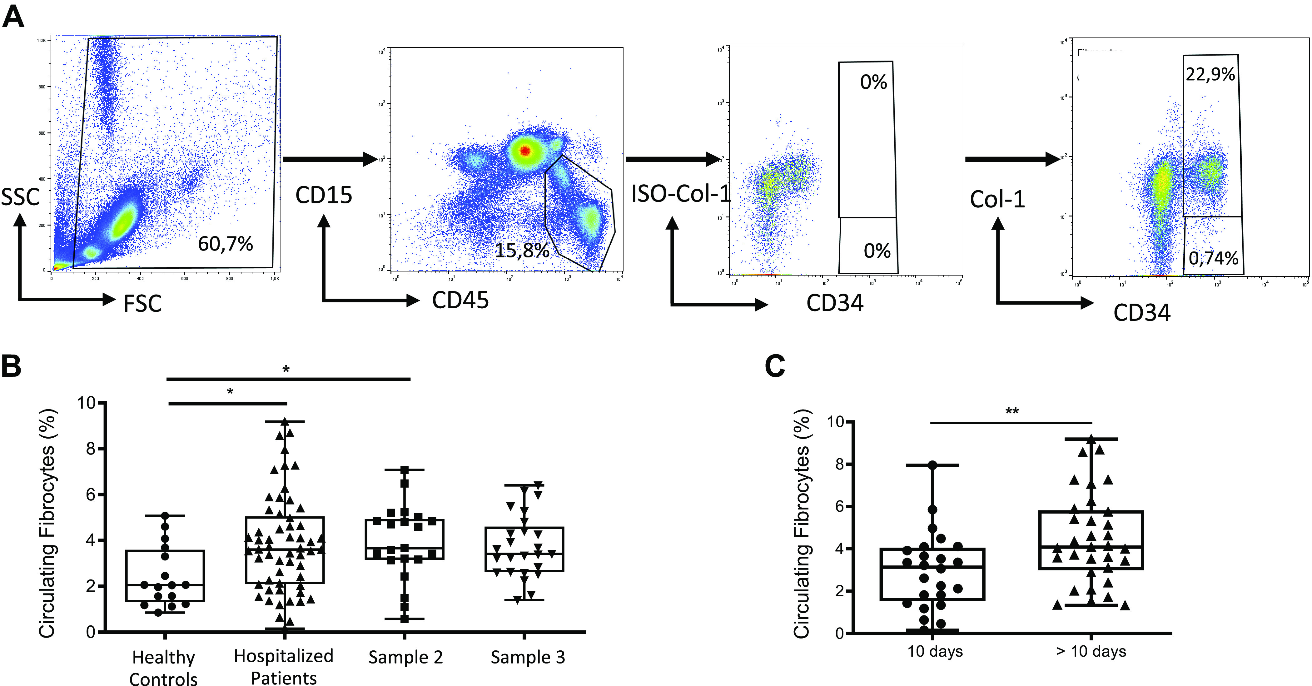
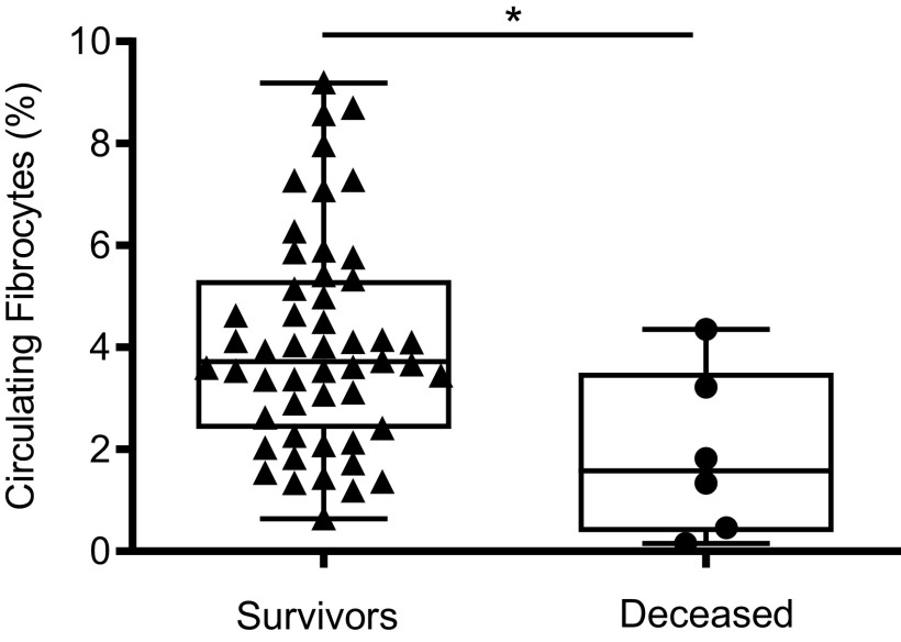
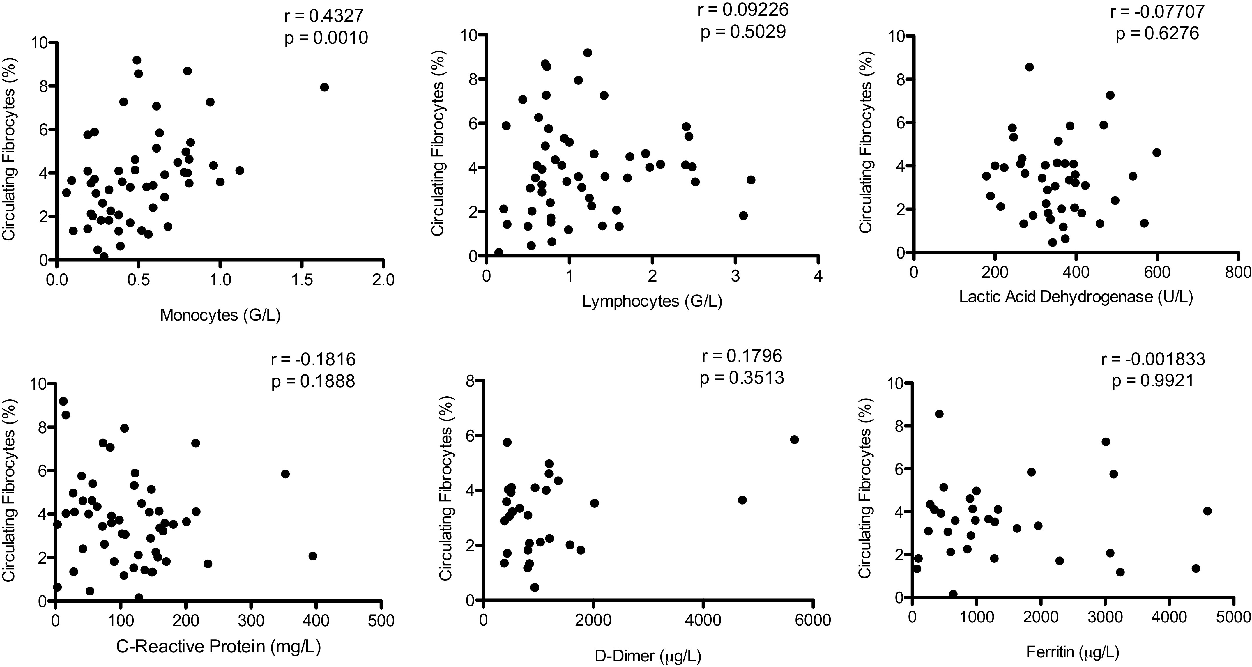
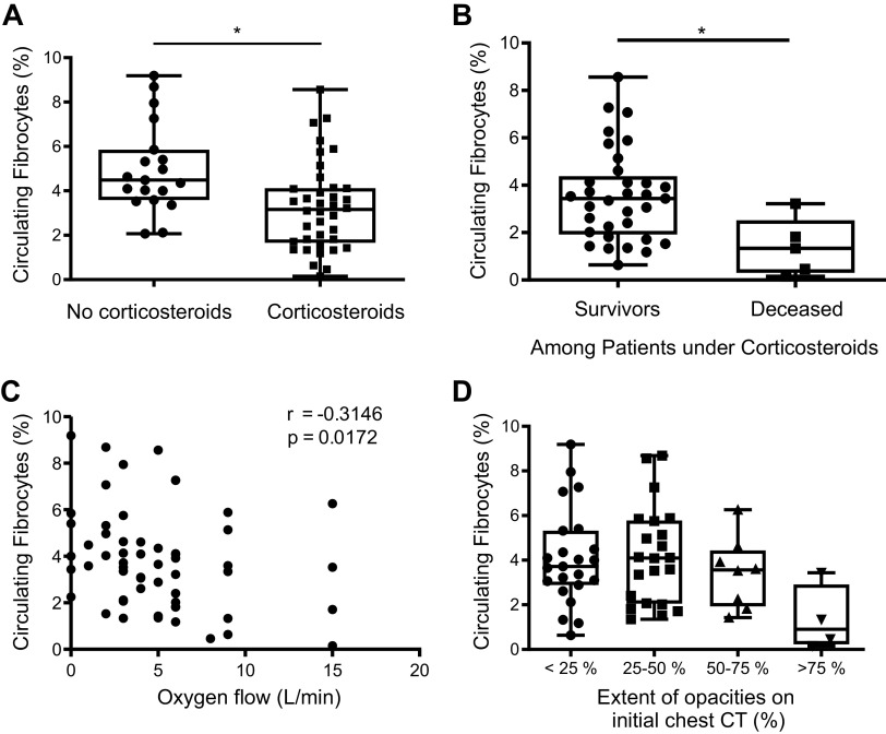
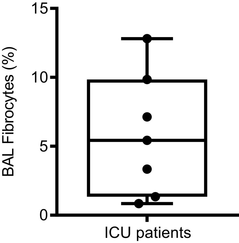
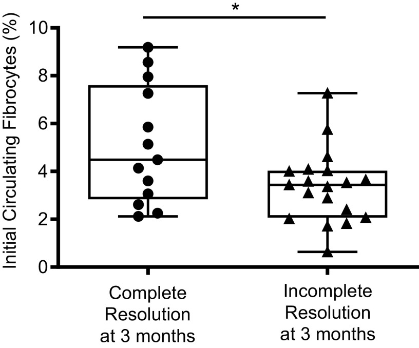
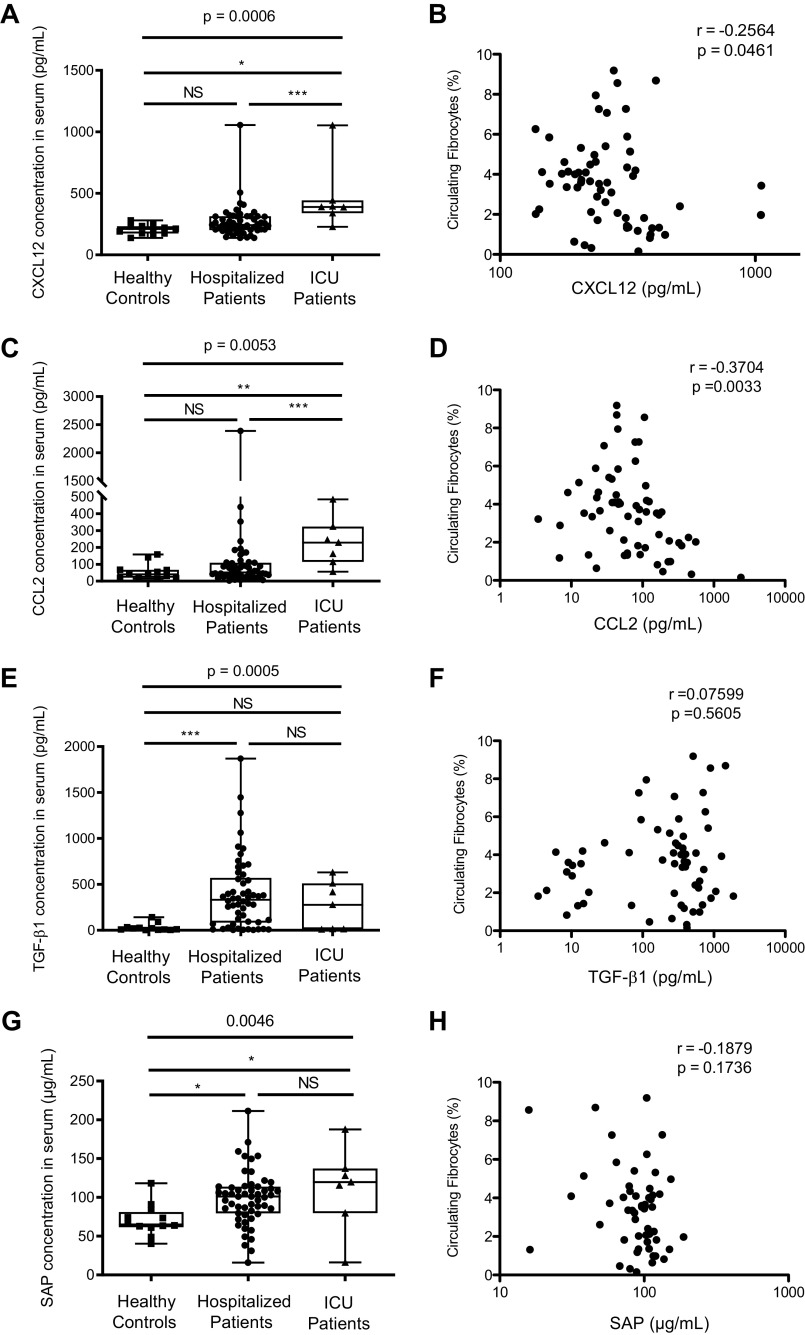
Increased blood fibrocytes are associated with a poor prognosis in fibrotic lung diseases. We aimed to determine whether the percentage of circulating fibrocytes could be predictive of severity and prognosis during coronavirus disease 2019 (COVID-19) pneumonia. Blood fibrocytes were quantified by flow cytometry as CD45+/CD15-/CD34+/collagen-1+ cells in patients hospitalized for COVID-19 pneumonia. In a subgroup of patients admitted in an intensive care unit (ICU), fibrocytes were quantified in blood and bronchoalveolar lavage (BAL). Serum amyloid P (SAP), transforming growth factor-β1 (TGF-β1), CXCL12, CCL2, and FGF2 concentrations were measured. We included 57 patients in the hospitalized group (median age = 59 yr [23-87]) and 16 individuals as healthy controls. The median percentage of circulating fibrocytes was higher in the patients compared with the controls (3.6% [0.2-9.2] vs. 2.1% [0.9-5.1], P = 0.04). Blood fibrocyte count was lower in the six patients who died compared with the survivors (1.6% [0.2-4.4] vs. 3.7% [0.6-9.2], P = 0.02). Initial fibrocyte count was higher in patients showing a complete lung computed tomography (CT) resolution at 3 mo. Circulating fibrocyte count was decreased in the ICU group (0.8% [0.1-2.0]), whereas BAL fibrocyte count was 6.7% (2.2-15.4). Serum SAP and TGF-β1 concentrations were increased in hospitalized patients. SAP was also increased in ICU patients. CXCL12 and CCL2 were increased in ICU patients and negatively correlated with circulating fibrocyte count. We conclude that circulating fibrocytes were increased in patients hospitalized for COVID-19 pneumonia, and a lower fibrocyte count was associated with an increased risk of death and a slower resolution of lung CT opacities.

Keywords: COVID-19 pneumonia; fibrocyte; prognosis.


Conflict of interest statement

B. Crestani is the principal investigator of the NINTECOR trial (NCT04541680) (Nintedanib for the Treatment of SARS-Cov-2 Induced Pulmonary Fibrosis). None of the other authors has any conflicts of interest, financial or otherwise, to disclose.

Figures

Figure 1.
Figure 1.
Circulating fibrocytes in hospitalized patients. A: gating strategy for quantification of circulating fibrocytes in peripheral blood. Population was initially gated on the basis of the side scatter characteristics (SSCs), and debris were eliminated. Then, lymphocytes and monocytes were selected with gating of the CD45+ CD15 population (excluding granulocytes). Finally, fibrocytes were selected based on the expression of CD34 and collagen-I (CD34+ Col-I+). Collagen-I isotype (ISO-Col-I) was used as a fluorescence minus one (FMO) control for the gating of Col-I+ population. B: blood circulating fibrocyte count was higher in patients hospitalized for COVID-19 pneumonia as compared with healthy controls. No significant difference was detected in repeated blood samples. A second blood sample was obtained on day 7 after the first sample in 21 patients (sample 2). A third sample was obtained on day 90 in 25 patients (sample 3). Dot box-and-whisker plots with median showing the percentage of circulating fibrocytes among live mononuclear leukocytes in healthy controls (n = 16) and hospitalized patients with COVID-19 (n = 57). Kruskal–Wallis test, *P = 0.03. C: blood circulating fibrocyte count was higher in samples collected >10 days after symptom onset. Dot box-and-whisker plots with median showing the percentage of circulating fibrocytes among live mononuclear leukocytes in hospitalized patients with COVID-19 sampled >10 days (n = 33) and ≤10 days after symptom onset (n = 24). Mann–Whitney U test, ** P ≤ 0.01.
Figure 2.
Figure 2.
Blood fibrocyte count was decreased in patients who died during hospitalization as compared with survivors. Dot box-and-whisker plots with median showing the percentage of circulating fibrocytes among live mononuclear leukocytes in survivors (n = 51) and in patients who died (n = 6). Mann–Whitney U test, *P ≤ 0.05.
Figure 3.
Figure 3.
Circulating fibrocyte count was positively correlated with blood monocyte count but was not correlated to biological markers associated with COVID-19 severity [lymphocyte count, lactate dehydrogenase (LDH), C-reactive protein (CRP), d-dimer, and ferritin]. Spearman rank-order correlation coefficient.
Figure 4.
Figure 4.
Circulating fibrocytes and severity. A: blood fibrocyte count was decreased in patients receiving corticosteroids for the treatment of COVID-19 before sample collection. Dot box-and-whisker plots with median showing the percentage of circulating fibrocytes among live mononuclear leukocytes in hospitalized patients treated with corticosteroids at the time of sampling (n = 38) and patients who were not treated (n = 19). Mann–Whitney U test, * P ≤ 0.05. B: blood fibrocyte count was lower in deceased patients as compared with survivors among hospitalized patients receiving corticosteroids for the treatment of COVID-19. Dot box-and-whisker plots with median showing the percentage of circulating fibrocytes among live mononuclear leukocytes in hospitalized patients treated with corticosteroids at the time of sampling (survivors, n = 33; deceased, n = 5). Mann–Whitney U test, *P ≤ 0.05. C: blood fibrocyte count was negatively correlated with oxygen flow in hospitalized patients with COVID-19. Spearman rank-order correlation coefficient. D: blood fibrocyte count was decreased when extension of pneumonia on computed tomography (CT) scan was >75%. Dot box-and-whisker plots with median showing the percentage of circulating fibrocytes among live mononuclear leukocytes according to the extension of the lesions on CT scan (<25%: n = 23; 25%–50%: n = 22; 50%–75%: n = 8; and >75%: n = 4). Kruskal–Wallis test, P = 0.07.
Figure 5.
Figure 5.
Percentage of fibrocytes in bronchoalveolar lavage (BAL) from patients with COVID-19 hospitalized in an intensive care unit (ICU). Dot box-and-whisker plots with median showing the percentage of fibrocytes among live mononuclear leukocytes in BAL of COVID-19 ICU patients (n = 7).
Figure 6.
Figure 6.
A higher initial circulating fibrocyte count was associated with a complete computed tomography (CT) resolution after 3-mo follow-up. Dot box-and-whisker plots with median showing the percentage of circulating fibrocytes among live mononuclear leukocytes in healthy controls (n = 16), patients with COVID-19 showing a complete CT resolution (n = 13), and patients with COVID-19 showing an incomplete CT resolution (n = 19). Mann–Whitney U test, * P ≤ 0.05.
Figure 7.
Figure 7.
Serum concentration of cytokines involved in fibrocyte recruitment and differentiation. A: CXCL12 concentration was significantly increased in intensive care unit (ICU) patients as compared with hospitalized patients and healthy controls. Dot box-and-whisker plots with median range showing the concentration of CXCL12 in healthy controls (n = 12), hospitalized patients (n = 54), and ICU patients (n = 7). Kruskal–Wallis test, P = 0.0006. B: CXCL12 concentration was negatively correlated with circulating fibrocytes count in hospitalized and ICU patients. CXCL12 concentration and percentage of circulating fibrocytes were measured in blood of hospitalized (n = 54) and ICU patients (n = 7). Spearman rank-order correlation coefficient. C: CCL2 concentration was increased in ICU patients as compared with hospitalized patients and healthy controls. Dot box-and-whisker plots with median range showing the concentration of CCL2 in healthy controls (n = 12), hospitalized patients (n = 54), and ICU patients (n = 7). Kruskal–Wallis test, P = 0.0053. D: CCL2 concentration was negatively correlated with circulating fibrocyte count in hospitalized and ICU patients. CCL2 concentration and percentage of circulating fibrocytes were measured in blood of hospitalized patients (n = 54) and ICU patients (n = 7). Spearman rank-order correlation coefficient. E: TGF-β1 concentration was increased in hospitalized patients with COVID-19 as compared with healthy controls. Dot box-and-whisker plots with median range showing the concentration of TGF-β1 in healthy controls (n = 12), hospitalized patients (n = 54), and ICU patients (n = 7). Kruskal–Wallis test, P = 0.0005. F: no correlation was found between TGF-β1 concentration in serum and circulating fibrocyte count in hospitalized and ICU patients. TGF-β1 concentration and percentage of circulating fibrocytes were measured in blood of hospitalized (n = 54) and ICU patients (n = 7). Spearman rank-order correlation coefficient. G: serum amyloid protein (SAP) concentration was increased in hospitalized patients with COVID-19 as compared with healthy controls. Dot box-and-whisker plots with median range showing the concentration of SAP in healthy controls (n = 12), hospitalized patients (n = 54), and ICU patients (n = 7). Kruskal–Wallis test, P = 0.046. H: no correlation was found between SAP concentration and circulating fibrocyte count in hospitalized and ICU patients. SAP concentration and percentage of circulating fibrocytes were measured in blood of hospitalized (n = 54) and ICU patients (n = 7). Spearman rank-order correlation coefficient. *P < 0.05; **P < 0.01; ***P < 0.001.
Figure 8.
Figure 8.
Detection of fibrocytes in COVID-19 lung samples using hematopoietic (CD45) and mesenchymal markers (vimentin) by fluorescence microscopy. Frozen lung sections from patients with COVID-19 were co-immunostained using CD45 and vimentin. DAPI was used for nuclei staining. Arrows indicate CD45 and vimentin double-positive cells. Scale bar: 50 µm. Patient 1 developed symptomatic COVID-19 pneumonia 2 days after bullous emphysema surgery, with RT-PCR positive for COVID-19 on the lung sample. Patient 2 underwent lung cancer surgery 4 wk after first symptoms of COVID-19 pneumonia.

References

    1. Huang C, Wang Y, Li X, Ren L, Zhao J, Hu Y, Zhang L, Fan G, Xu J, Gu X, Cheng Z, Yu T, Xia J, Wei Y, Wu W, Xie X, Yin W, Li H, Liu M, Xiao Y, Gao H, Guo L, Xie J, Wang G, Jiang R, Gao Z, Jin Q, Wang J, Cao B. Clinical features of patients infected with 2019 novel coronavirus in Wuhan, China. Lancet 395: 497–506, 2020. doi:10.1016/S0140-6736(20)30183-5. - DOI - PMC - PubMed
    1. Elezkurtaj S, Greuel S, Ihlow J, Michaelis EG, Bischoff P, Kunze CA, Sinn BV, Gerhold M, Hauptmann K, Ingold-Heppner B, Miller F, Herbst H, Corman VM, Martin H, Radbruch H, Heppner FL, Horst D. Causes of death and comorbidities in hospitalized patients with COVID-19. Sci Rep 11: 4263, 2021. doi:10.1038/s41598-021-82862-5. - DOI - PMC - PubMed
    1. Bourgonje AR, Abdulle AE, Timens W, Hillebrands J, Navis GJ, Gordijn SJ, Bolling MC, Dijkstra G, Voors AA, Osterhaus AD, Voort PH, Mulder DJ, Goor H. Angiotensin‐converting enzyme 2 (ACE2), SARS‐CoV‐2 and the pathophysiology of coronavirus disease 2019 (COVID‐19). J Pathol 251: 228–248, 2020. doi:10.1002/path.5471. - DOI - PMC - PubMed
    1. Hariri LP, North CM, Shih AR, Israel RA, Maley JH, Villalba JA, Vinarsky V, Rubin J, Okin DA, Sclafani A, Alladina JW, Griffith JW, Gillette MA, Raz Y, Richards CJ, Wong AK, Ly A, Hung YP, Chivukula RR, Petri CR, Calhoun TF, Brenner LN, Hibbert KA, Medoff BD, Hardin CC, Stone JR, Mino-Kenudson M. Lung histopathology in coronavirus disease 2019 as compared with severe acute respiratory syndrome and H1N1 influenza: a systematic review. Chest 159: 73–84, 2021. doi:10.1016/j.chest.2020.09.259. - DOI - PMC - PubMed
    1. Fox SE, Akmatbekov A, Harbert JL, Li G, Brown JQ, Vander Heide RS. Pulmonary and cardiac pathology in Covid-19: the first autopsy series from New Orleans. medRxiv 2020.04.06.20050575, 2020. doi:10.1101/2020.04.06.20050575. - DOI

Publication types