. 2021 Sep 8;19(1):135.
doi: 10.1186/s12958-021-00821-1.

Irisin alleviates obesity-related spermatogenesis dysfunction via the regulation of the AMPKα signalling pathway

Affiliations

Irisin alleviates obesity-related spermatogenesis dysfunction via the regulation of the AMPKα signalling pathway

Yang Mu et al. Reprod Biol Endocrinol. .

Abstract

Background: Infertility is a common complication in obese men. Oxidative stress and testicular apoptosis play critical roles in obesity-induced spermatogenesis dysfunction. It has been reported that irisin, an exercise-induced myokine, may attenuate oxidative damage and testicular apoptosis in several diseases; however, its role in obesity-induced spermatogenesis dysfunction remains unclear. The purpose of this study was to investigate the role and underlying mechanism of irisin in obesity-induced dysfunction of spermatogenesis.

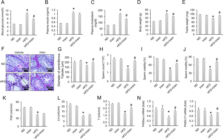
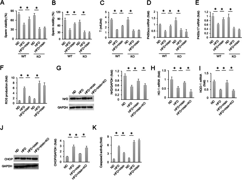
Methods: Male mice were fed a high-fat diet (HFD) for 24 weeks to establish a model of obesity-induced spermatogenesis dysfunction. To explore the effects of irisin, mice were subcutaneously infused with recombinant irisin for 8 weeks beginning at 16 weeks after starting a HFD. To confirm the role of AMP-activated protein kinase α (AMPKα), AMPKα-deficient mice were used.

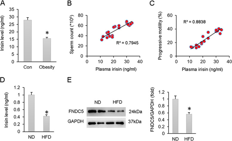
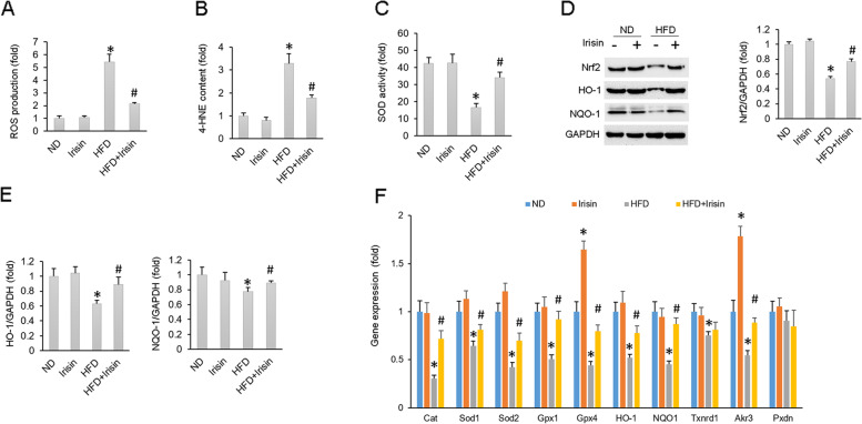
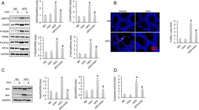
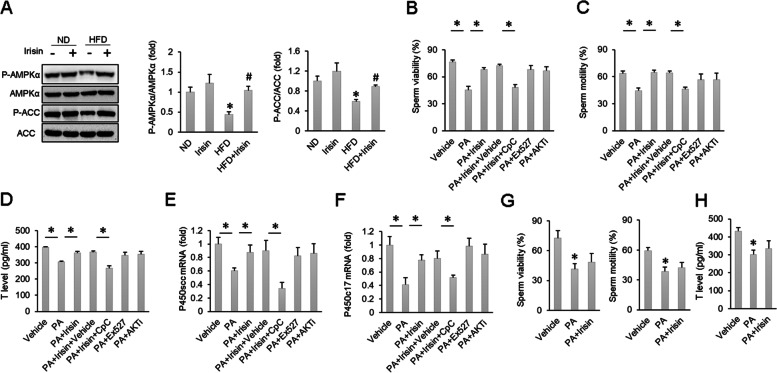
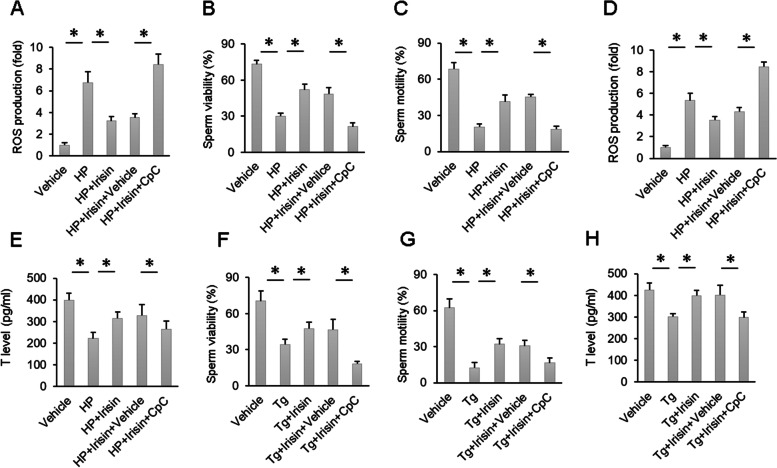
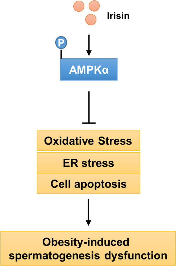
Results: The data showed decreased serum irisin levels in obese patients, which was negatively correlated with sperm count and progressive motility. Irisin was downregulated in the plasma and testes of obese mice. Supplementation with irisin protected against HFD-induced spermatogenesis dysfunction and increased testosterone levels in mice. HFD-induced oxidative stress, endoplasmic reticulum (ER) stress and testicular apoptosis were largely attenuated by irisin treatment. Mechanistically, we identified that irisin activated the AMPKα signalling pathway. With AMPKα depletion, we found that the protective effects of irisin on spermatogenesis dysfunction were abolished in vivo and in vitro.

Conclusions: In conclusion, we found that irisin alleviated obesity-related spermatogenesis dysfunction via activation of the AMPKα signalling pathway. Based on these findings, we hypothesized that irisin is a potential therapeutic agent against obesity-related spermatogenesis dysfunction.

Keywords: AMPKα; HFD; Irisin; Spermatogenesis dysfunction.


Conflict of interest statement

None declared.

Figures

Fig. 1
Fig. 1
Irisin expression is downregulated by obesity. A The serum irisin levels of obese and control humans (Con: n = 10; Obesity: n = 9). B Correlation between sperm count and plasma irisin level (Con: n = 10; Obesity: n = 9). C Correlation between progressive sperm rate and plasma irisin level (Con: n = 10; Obesity: n = 9). D Serum irisin level of HFD mice and ND mice (n = 6). E FNDC5 expression in the testes of HFD mice and ND mice (n = 6). Data are presented as the mean ± SEM. *P < 0.05 vs. the matched control
Fig. 2
Fig. 2
HFD-induced male spermatogenesis dysfunction was improved by irisin treatment. A Blood glucose level (n = 12). B Plasma insulin level (n = 12). C Plasma cholesterol level (n = 12). D-E Body weight and testes weight of the indicated groups (n = 12). F HE staining of mice testes. G Statistical analysis of the diameter of seminiferous tubules in the four groups (n = 6). H-J Sperm count, sperm viability and sperm motility (n = 6). K-M Serum FSH, LH and testosterone levels of the indicated groups (n = 6). N The mRNA expression of P450scc and P450c17 (n = 6). Data are presented as the mean ± SEM. *P < 0.05 vs. the matched control. Atrophied seminiferous tubules are marked with a double-head arrow. Impairment of the blood-testis barrier in the testis is marked with an arrowhead
Fig. 3
Fig. 3
Oxidative stress was suppressed by irisin. A Testicular ROS production (n = 6). B Testicular A-HNE content (n = 6). C SOD activity in mice testes (n = 6). D-E Protein levels of Nrf2, HO-1 and NQO-1 (n = 6). F Oxidative stress-related gene expression in mouse testes (n = 6). Data are presented as the mean ± SEM. *P < 0.05 vs. the matched control
Fig. 4
Fig. 4
ER stress and cell apoptosis were attenuated by irisin. A Protein level of ER stress related markers (n = 6). B TUNEL staining of mice testes (n = 5). C Protein levels of Bim and Bax in mouse testes (n = 6). D Testicular caspase 3 activity (n = 6). Data are presented as the mean ± SEM. *P < 0.05 vs. the matched control
Fig. 5
Fig. 5
The protective effects of irisin on HFD-induced spermatogenesis dysfunction were abolished after AMPK inhibition. A Protein level of AMPK and ACC (n = 6). B-C Sperm viability and sperm motility of the indicated groups (n = 6). D-F T level and mRNA expression of P450scc and P450c17 (n = 6). G-H Sperm viability, sperm motility and T levels after AMPK knockout (n = 6). Data are presented as the mean ± SEM. *P < 0.05 vs. the matched control
Fig. 6
Fig. 6
HP- and Tg-induced sperm damage and ROS production and a reduction in T levels were ameliorated by AMPK inhibition. A ROS production of sperm in the indicated groups (n = 6). B-C Sperm viability and sperm motility of the indicated groups (n = 6). D ROS production of Leydig cells in the indicated groups (n = 6). E T production of Leydig cells in the indicated groups (n = 6). F-G Sperm viability and sperm motility of the indicated groups (n = 6). H T production of Leydig cells in the indicated groups (n = 6). Data are presented as the mean ± SEM. *P < 0.05 vs. the matched control
Fig. 7
Fig. 7
The protective effects of irisin on spermatogenesis function were abolished after AMPK knockout. A-B Sperm viability and sperm motility of the indicated groups (n = 6). C-E T level and mRNA expression of P450scc and P450c17 (n = 6). F ROS production of the indicated groups (n = 6). G Nrf2 expression in mice testes (n = 6). H-I mRNA levels of HO-1 and NQO-1 in mouse testes (n = 6). J CHOP expression in mice testes (n = 6). K Testicular Caspase-3 activity of the indicated groups (n = 6). Data are presented as the mean ± SEM. *P < 0.05 vs. the matched control
Fig. 8
Fig. 8
Schematic diagram of the molecular mechanisms underlying irisin-mediated protection of spermatogenic functions in mice

Similar articles

Cited by

References

    1. Ombelet W, Cooke I, Dyer S, Serour G, Devroey P. Infertility and the provision of infertility medical services in developing countries. Hum Reprod Update. 2008;14(6):605–621. doi: 10.1093/humupd/dmn042. - DOI - PMC - PubMed
    1. Jarow JP, Sharlip ID, Belker AM, Lipshultz LI, Sigman M, Thomas AJ, Schlegel PN, Howards SS, Nehra A, Damewood MD, Overstreet JW, Sadovsky R. Best practice policies for male infertility. J Urol. 2002;167(5):2138–2144. doi: 10.1016/S0022-5347(05)65109-9. - DOI - PubMed
    1. Best D, Bhattacharya S. Obesity and fertility. Horm Mol Biol Clin Investig. 2015;24(1):5–10. - PubMed
    1. Hammoud A, Carrell DT, Meikle AW, Xin Y, Hunt SC, Adams TD, Gibson M. An aromatase polymorphism modulates the relationship between weight and estradiol levels in obese men. Fertil Steril. 2010;94(5):1734–1738. doi: 10.1016/j.fertnstert.2009.10.037. - DOI - PMC - PubMed
    1. Aitken J, Fisher H. Reactive oxygen species generation and human spermatozoa: the balance of benefit and risk. Bioessays. 1994;16(4):259–267. doi: 10.1002/bies.950160409. - DOI - PubMed

MeSH terms