

Histological differentiation impacts the tumor immune microenvironment in gastric carcinoma: Relation to the immune cycle
- PMID: 34497449
- PMCID: PMC8384749
- DOI: 10.3748/wjg.v27.i31.5259
Histological differentiation impacts the tumor immune microenvironment in gastric carcinoma: Relation to the immune cycle
Abstract
Background: Various histological types of gastric carcinomas (GCs) differ in terms of their pathogenesis and their preexisting background, both of which could impact the tumor immune microenvironment (TIME). However, the current understanding of the immune contexture of GC is far from complete.
Aim: To clarify the tumor-host immune interplay through histopathological features and the tumor immune cycle concept.
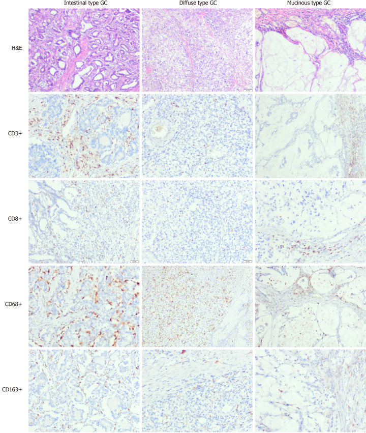
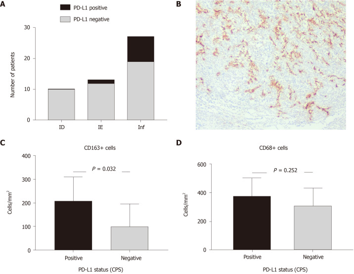
Methods: In total, 50 GC cases were examined (15 cases of diffuse GC, 31 patients with intestinal-type GC and 4 cases of mucinous GC). The immunophenotype of GC was assessed and classified as immune desert (ID), immune excluded (IE) or inflamed (Inf) according to CD8+ cell count and spatial pattern. In addition, CD68+ and CD163+ macrophages and programmed death-ligand 1 (PD-L1) expression were estimated.
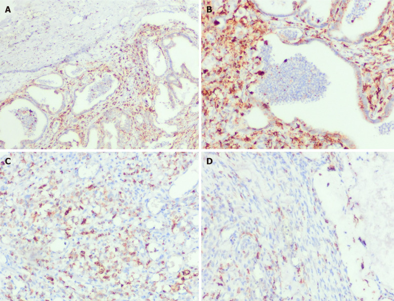
Results: We found that GCs with different histological differentiation demonstrated distinct immune contexture. Most intestinal-type GCs had inflamed TIMEs rich in both CD8+ cells and macrophages. In contrast, more aggressive diffuse-type GC more often possessed ID characteristics with few CD8+ lymphocytes but abundant CD68+ macrophages, while mucinous GC had an IE-TIME with a prevalence of CD68+ macrophages and CD8+ lymphocytes in the peritumor stroma. PD-L1 expression prevailed mostly in intestinal-type Inf-GC, with numerous CD163+ cells observed. Therefore, GCs of different histological patterns have specific mechanisms of immune escape. While intestinal-type GC was more often related to PD-L1 expression, diffuse and mucinous GCs possessing more aggressive behavior demonstrated low immunogenicity and a lack of tumor antigen recognition or immune cell recruitment into the tumor clusters.
Conclusion: These data help to clarify the links between tumor histogenesis and immunogenicity for a better understanding of GC biology and more tailored patient management.
Keywords: Gastric carcinoma; Tumor associated macrophages; Tumor immune microenvironment; Tumor infiltrating lymphocytes.
©The Author(s) 2021. Published by Baishideng Publishing Group Inc. All rights reserved.
Conflict of interest statement
Conflict-of-interest statement: We have no financial relationships to disclose.
Figures
References
-
- Siegel RL, Miller KD, Jemal A. Cancer statistics, 2019. CA Cancer J Clin. 2019;69:7–34. - PubMed
-
- Ji X, Bu ZD, Yan Y, Li ZY, Wu AW, Zhang LH, Zhang J, Wu XJ, Zong XL, Li SX, Shan F, Jia ZY, Ji JF. The 8th edition of the American Joint Committee on Cancer tumor-node-metastasis staging system for gastric cancer is superior to the 7th edition: results from a Chinese mono-institutional study of 1663 patients. Gastric Cancer. 2018;21:643–652. - PMC - PubMed
-
- Lauren P. The two histological main types of gastric carcinoma: Diffuse and so-called intestinal-type carcinoma. An attempt at a histo-clinical classification. Acta Pathol Microbiol Scand. 1965;64:31–49. - PubMed
-
- Chen YC, Fang WL, Wang RF, Liu CA, Yang MH, Lo SS, Wu CW, Li AF, Shyr YM, Huang KH. Clinicopathological Variation of Lauren Classification in Gastric Cancer. Pathol Oncol Res. 2016;22:197–202. - PubMed
-
- Giraldo NA, Peske JD, Sautès-Fridman C, Fridman WH. Integrating histopathology, immune biomarkers, and molecular subgroups in solid cancer: the next step in precision oncology. Virchows Arch. 2019;474:463–474. - PubMed
MeSH terms
Substances
LinkOut - more resources
Full Text Sources
Medical
Research Materials
Miscellaneous
