. 2021 Aug 23:15:701308.
doi: 10.3389/fnins.2021.701308. eCollection 2021.

Contribution of Tissue Inflammation and Blood-Brain Barrier Disruption to Brain Softening in a Mouse Model of Multiple Sclerosis

Affiliations

Contribution of Tissue Inflammation and Blood-Brain Barrier Disruption to Brain Softening in a Mouse Model of Multiple Sclerosis

Rafaela Vieira Silva et al. Front Neurosci. .

Abstract

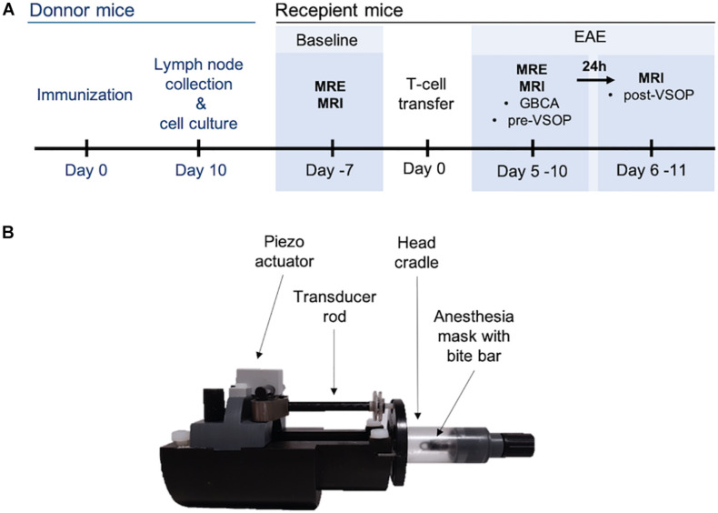
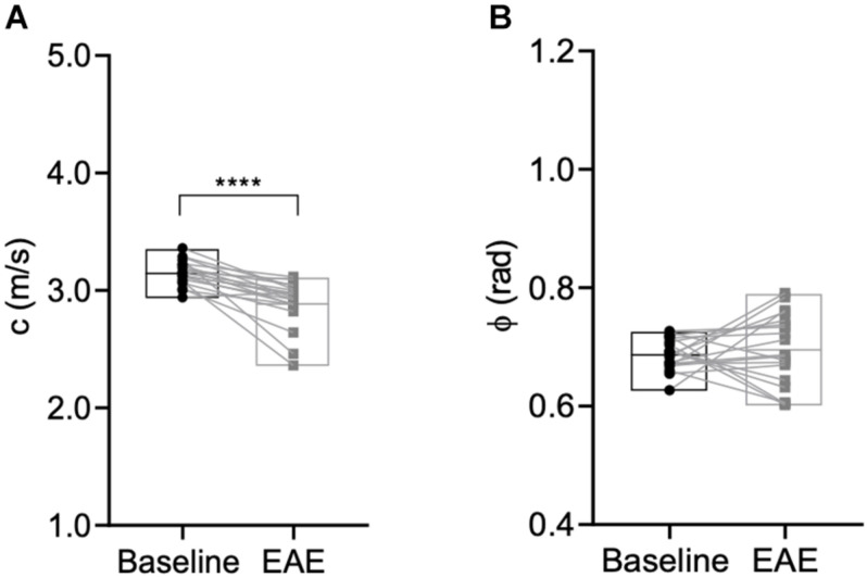
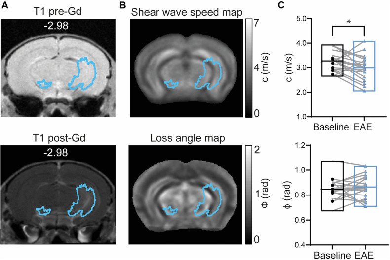
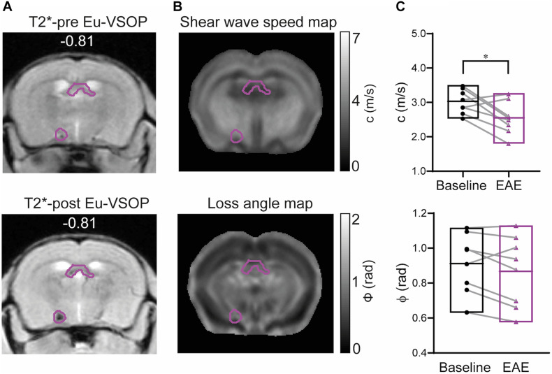
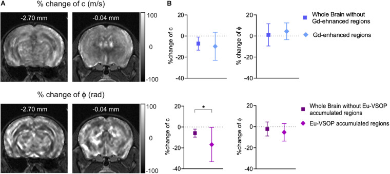
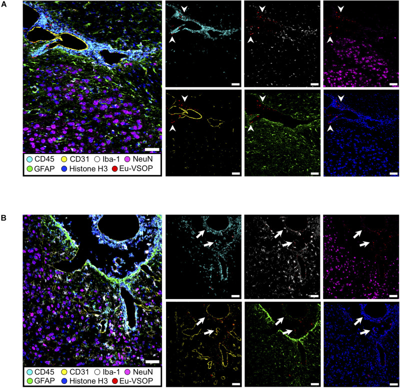
Neuroinflammatory processes occurring during multiple sclerosis cause disseminated softening of brain tissue, as quantified by in vivo magnetic resonance elastography (MRE). However, inflammation-mediated tissue alterations underlying the mechanical integrity of the brain remain unclear. We previously showed that blood-brain barrier (BBB) disruption visualized by MRI using gadolinium-based contrast agent (GBCA) does not correlate with tissue softening in active experimental autoimmune encephalomyelitis (EAE). However, it is unknown how confined BBB changes and other inflammatory processes may determine local elasticity changes. Therefore, we aim to elucidate which inflammatory hallmarks are determinant for local viscoelastic changes observed in EAE brains. Hence, novel multifrequency MRE was applied in combination with GBCA-based MRI or very small superparamagnetic iron oxide particles (VSOPs) in female SJL mice with induced adoptive transfer EAE (n = 21). VSOPs were doped with europium (Eu-VSOPs) to facilitate the post-mortem analysis. Accumulation of Eu-VSOPs, which was previously demonstrated to be sensitive to immune cell infiltration and ECM remodeling, was also found to be independent of GBCA enhancement. Following registration to a reference brain atlas, viscoelastic properties of the whole brain and areas visualized by either Gd or VSOP were quantified. MRE revealed marked disseminated softening across the whole brain in mice with established EAE (baseline: 3.1 ± 0.1 m/s vs. EAE: 2.9 ± 0.2 m/s, p < 0.0001). A similar degree of softening was observed in sites of GBCA enhancement i.e., mainly within cerebral cortex and brain stem (baseline: 3.3 ± 0.4 m/s vs. EAE: 3.0 ± 0.5 m/s, p = 0.018). However, locations in which only Eu-VSOP accumulated, mainly in fiber tracts (baseline: 3.0 ± 0.4 m/s vs. EAE: 2.6 ± 0.5 m/s, p = 0.023), softening was more pronounced when compared to non-hypointense areas (percent change of stiffness for Eu-VSOP accumulation: -16.81 ± 16.49% vs. for non-hypointense regions: -5.85 ± 3.81%, p = 0.048). Our findings suggest that multifrequency MRE is sensitive to differentiate between local inflammatory processes with a strong immune cell infiltrate that lead to VSOP accumulation, from disseminated inflammation and BBB leakage visualized by GBCA. These pathological events visualized by Eu-VSOP MRI and MRE may include gliosis, macrophage infiltration, alterations of endothelial matrix components, and/or extracellular matrix remodeling. MRE may therefore represent a promising imaging tool for non-invasive clinical assessment of different pathological aspects of neuroinflammation.

Keywords: BBB disruption; Eu-VSOP; experimental autoimmune encephalomyelitis; gadolinium; magnetic resonance elastography; multiple sclerosis; neuroinflammation.


Conflict of interest statement

The authors declare that the research was conducted in the absence of any commercial or financial relationships that could be construed as a potential conflict of interest.

Figures

FIGURE 1
FIGURE 1
Experimental set-up. (A) Timeline depicting induction of adoptive transfer EAE and in vivo scans at baseline, before immunization, and after establishment of EAE. (B) Custom-made animal holder. A piezo actuator generates the mechanical waves, which are transmitted via the transducer to the head cradle and into the skull of the mouse.
FIGURE 2
FIGURE 2
Representative wave images. Propagation of shear waves through the mouse brain after k-MDEV inversion (1 slice, 1 component, 5 frequencies, and no directional filters applied). The image was taken from a mouse after immunization.
FIGURE 3
FIGURE 3
Percentage incidence maps for Gd-enhancement and Eu-VSOP accumulation during EAE. (A) Incidence (% of mice affected) of gadolinium distribution across the brain, displaying large diffuse lesions along the cerebral cortex, cerebral nuclei, brain stem, midbrain and fiber tracts (n = 19). (B) Conversely, Eu-VSOP distribution appears to be characterized by smaller and focal areas of accumulation in the cerebral cortex, cerebral nuclei, brain stem, midbrain and fiber tracts (n = 11).
FIGURE 4
FIGURE 4
Global brain softening due to inflammation. (A) Whole brain stiffness is reduced during inflammation (p < 0.0001, Wilcoxon matched-pairs signed rank test), whereas (B) fluidity is unaffected (p = 0.6070, paired t-test). n = 19; mean, min/max. ****p ≤ 0.0001.
FIGURE 5
FIGURE 5
Brain mechanical properties in Gd-enhanced lesions during EAE. (A) Representative T1-weighted image acquired before (left) and after GBCA (right) injection (corresponding GBCA mask overlaid) shows Gd-enhancing lesions near the left hippocampal artery. (B) GBCA mask overlaid on averaged MRE maps of stiffness (shear wave speed) and fluidity (loss angle). (C) Stiffness represented by shear wave speed (c) was reduced during EAE (p = 0.0183; paired t-test) whereas tissue fluidity (ϕ) was unchanged (p = 0.3683; paired t test). n = 19; mean, min/max; *<0.05. Of note, ventricles are excluded from our analysis since MRE cannot be performed in fluid compartments.
FIGURE 6
FIGURE 6
Brain mechanical properties of areas accumulating Eu-VSOPs during EAE. (A) Exemplary image of a mask comprising Eu-VSOP accumulation overlaid on T2*-weighted image prior (left) and post Eu-VSOP (right) injection. (B) Shear wave speed map (c in m/s) and loss angle map (ϕ in rad). (C) There is a significant reduction in stiffness in sites with Eu-VSOP accumulation compared to non-hypointense areas (p = 0.0235, paired t-test). Fluidity of brain tissue in these areas, marked by ϕ, is not affected (p = 0.1168, paired t-test). n = 8, mean, min/max; *<0.05. Of note, ventricles are excluded from our analysis since MRE cannot be performed in fluid compartments.
FIGURE 7
FIGURE 7
MRE is sensitive to stiffness changes in sites of Eu-VSOP accumulation. (A) Representative percentage change maps of c- and ϕ-map. (B) Percentage change in areas with Gd enhancement (within cerebral cortex, cerebral nuclei, brain stem, midbrain and fiber tracts) compared to non-enhancing areas revealed no significant difference (top), for c in m/s p = 0.3321 (Wilcoxon matched-pairs rank sum test), for ϕ in rad p = 0.1246 (paired t-test), n = 19; percentage change of region with Eu-VSOP accumulation (within cerebral cortex, cerebral nuclei, brain stem, midbrain and fiber tracts) (bottom) compared to non-hypointense areas showed a significant difference for c (p = 0.0483, paired t-test, mean ± SD, n = 8, *<0.05) whereas ϕ was not affected (p = 0.2889, paired t-test, mean ± SD n = 8, *<0.05). Percentage change maps are based on the percentage difference from peak to baseline.
FIGURE 8
FIGURE 8
Visualization of brain inflammation induced by EAE using IMC. (A) Brain section of a healthy control (naïve) mouse at the third ventricles (left) and brain stem and hippocampal formation (right). (B) Extensive inflammation during EAE can be visualized by infiltration of CD45+ cells (cyan), astrogliosis (GFAP, green) and activated microglia (Iba-1, white) (white arrows). Neurons (NeuN—magenta), astrocytes (GFAP—green), endothelial cells (CD31—yellow), nuclei (histone H3—blue), microglia (Iba-1—white), leucocytes (CD45—cyan). Scale bar: 100 μm.
FIGURE 9
FIGURE 9
Histological visualization of Eu-VSOP distribution by IMC. (A) Magnetic particle accumulation in the perivascular space with strong leukocyte infiltrate (arrowheads) located between brain stem and hippocampal formation. (B) In the third ventricle, particles can be found in the choroid plexus and are associated with perivascular inflammation (arrows). Eu-VSOPs (red dots), neurons (NeuN—magenta), astrocytes (GFAP—green), endothelial cells (CD31—yellow), nuclei (histone H3—blue), microglia (Iba-1—white), leucocytes (CD45—cyan). Scale bar: 50 mm.

References

    1. Aime S., Caravan P. (2009). Biodistribution of gadolinium-based contrast agents, including gadolinium deposition. J. Magn. Reson. Imaging 30 1259–1267. 10.1002/jmri.21969 - DOI - PMC - PubMed
    1. Alvarez J. I., Cayrol R., Prat A. (2011). Disruption of central nervous system barriers in multiple sclerosis. Biochim. Biophys. Acta 1812 252–264. - PubMed
    1. Berndt D., Millward J. M., Schnorr J., Taupitz M., Stangl V., Paul F., et al. (2017). Inflammation-induced brain endothelial activation leads to uptake of electrostatically stabilized iron oxide nanoparticles via sulfated glycosaminoglycans. Nanomedicine 13 1411–1421. 10.1016/j.nano.2017.01.010 - DOI - PubMed
    1. Bertalan G., Guo J., Tzschätzsch H., Klein C., Barnhill E., Sack I., et al. (2019). Fast tomoelastography of the mouse brain by multifrequency single-shot MR elastography. Magn. Reson. Med. 81 2676–2687. 10.1002/mrm.27586 - DOI - PubMed
    1. Bertalan G., Klein C., Schreyer S., Steiner B., Kreft B., Tzschätzsch H., et al. (2020). Biomechanical properties of the hypoxic and dying brain quantified by magnetic resonance elastography. Acta Biomater. 101 395–402. 10.1016/j.actbio.2019.11.011 - DOI - PubMed

LinkOut - more resources