

An Inhibitory Medial Preoptic Circuit Mediates Innate Exploration
- PMID: 34497488
- PMCID: PMC8419349
- DOI: 10.3389/fnins.2021.716147
An Inhibitory Medial Preoptic Circuit Mediates Innate Exploration
Abstract
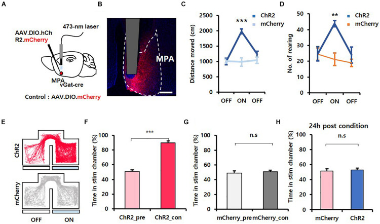
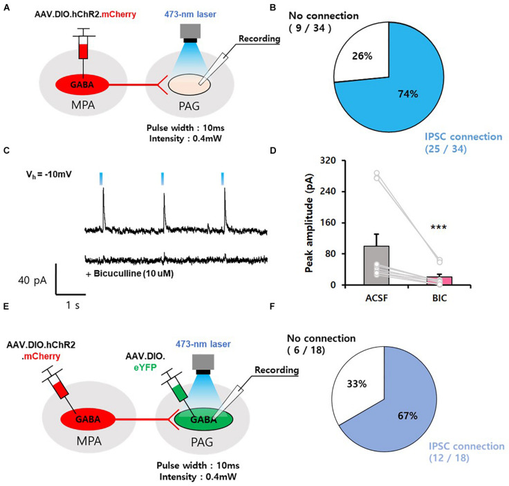
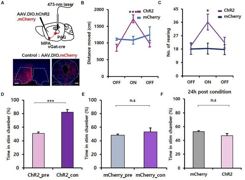
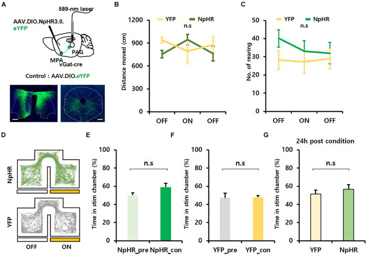
Animals have an innate motivation to explore objects and environments with unknown values. To this end, they need to activate neural pathways that enable exploration. Here, we reveal that photostimulation of a subset of medial preoptic area (MPA) neurons expressing the vesicular-GABA transporter gene (vgat+) and sending axonal projections to the ventrolateral periaqueductal gray (vPAG) increases exploration in a chamber but causes no place preference when tested there without photostimulation. Photoinhibition of MPAvgat-vPAG projections leads to no emotional changes as measured by normal activity in an open field assay. Electrophysiological recordings revealed that most GABAergic vPAG neurons are inhibited by MPAvgat neurons. In contrast to a previous report that suggested that MPAvgat-vPAG neurons may impart positive valence to induce place preference, our results suggest that these neurons can increase innate exploration.
Keywords: GABAergic neuron; exploration; medial preoptic area; periaqueductal gray; reinforcement.
Copyright © 2021 Ryoo, Park and Kim.
Conflict of interest statement
The authors declare that the research was conducted in the absence of any commercial or financial relationships that could be construed as a potential conflict of interest.
Figures
Similar articles
-
Exploration driven by a medial preoptic circuit facilitates fear extinction in mice.Commun Biol. 2023 Jan 27;6(1):106. doi: 10.1038/s42003-023-04442-9. Commun Biol. 2023. PMID: 36707677 Free PMC article.
-
Medial preoptic circuit induces hunting-like actions to target objects and prey.Nat Neurosci. 2018 Mar;21(3):364-372. doi: 10.1038/s41593-018-0072-x. Epub 2018 Jan 29. Nat Neurosci. 2018. PMID: 29379117
-
GABA neurons of the ventral periaqueductal gray area modulate behaviors associated with anxiety and conditioned fear.Brain Struct Funct. 2018 Nov;223(8):3787-3799. doi: 10.1007/s00429-018-1724-z. Epub 2018 Aug 4. Brain Struct Funct. 2018. PMID: 30076467
-
Maternal behaviour in lactating rats stimulates c-fos in glutamate decarboxylase-synthesizing neurons of the medial preoptic area, ventral bed nucleus of the stria terminalis, and ventrocaudal periaqueductal gray.Neuroscience. 2000;100(3):557-68. doi: 10.1016/s0306-4522(00)00287-6. Neuroscience. 2000. PMID: 11098119
-
AGRP Neurons Project to the Medial Preoptic Area and Modulate Maternal Nest-Building.J Neurosci. 2019 Jan 16;39(3):456-471. doi: 10.1523/JNEUROSCI.0958-18.2018. Epub 2018 Nov 20. J Neurosci. 2019. PMID: 30459220 Free PMC article.
Cited by
-
Exploration driven by a medial preoptic circuit facilitates fear extinction in mice.Commun Biol. 2023 Jan 27;6(1):106. doi: 10.1038/s42003-023-04442-9. Commun Biol. 2023. PMID: 36707677 Free PMC article.
-
Neural and Genetic Basis of Evasion, Approach and Predation.Mol Cells. 2022 Feb 28;45(2):93-97. doi: 10.14348/molcells.2022.2032. Mol Cells. 2022. PMID: 35236784 Free PMC article. Review.
-
Excitation-inhibition imbalance in medial preoptic area circuits underlies chronic stress-induced depression-like states.Nat Commun. 2024 Oct 3;15(1):8575. doi: 10.1038/s41467-024-52727-2. Nat Commun. 2024. PMID: 39362860 Free PMC article.
-
Hypothalamic control of innate social behaviors.Science. 2023 Oct 27;382(6669):399-404. doi: 10.1126/science.adh8489. Epub 2023 Oct 26. Science. 2023. PMID: 37883550 Free PMC article. Review.
-
Neurocircuitry of Predatory Hunting.Neurosci Bull. 2023 May;39(5):817-831. doi: 10.1007/s12264-022-01018-1. Epub 2023 Jan 27. Neurosci Bull. 2023. PMID: 36705845 Free PMC article. Review.
References
-
- Brown R. E., Corey S. C., Moore A. K. (1999). Differences in measures of exploration and fear in MHC-congenic C57BL/6J and B6-H-2K mice. Behav. Genet. 29 263–271.
-
- Dyne L. J., Hughes R. N. (1970). Effects of methylphenidate on activity and reactions to novelty in rats. Psychonomic Sci. 19 267–268. 10.3758/bf03328810 - DOI
LinkOut - more resources
Full Text Sources
Molecular Biology Databases