Review
. 2021 Aug 23:12:700220.
doi: 10.3389/fphys.2021.700220. eCollection 2021.

Angiotensin-Converting Enzyme 2 in the Pathogenesis of Renal Abnormalities Observed in COVID-19 Patients

Affiliations
Review

Angiotensin-Converting Enzyme 2 in the Pathogenesis of Renal Abnormalities Observed in COVID-19 Patients

Nayara Azinheira Nobrega Cruz et al. Front Physiol. .

Abstract

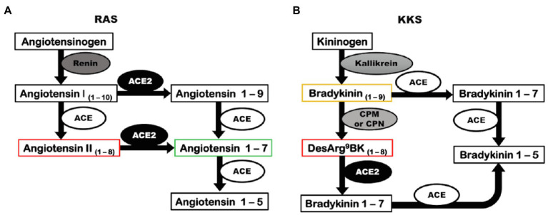
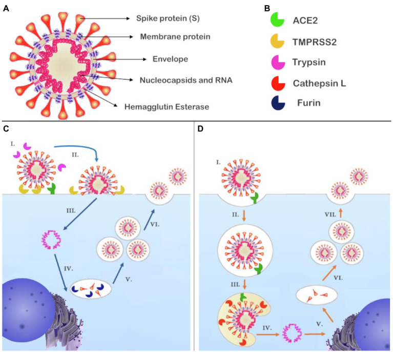
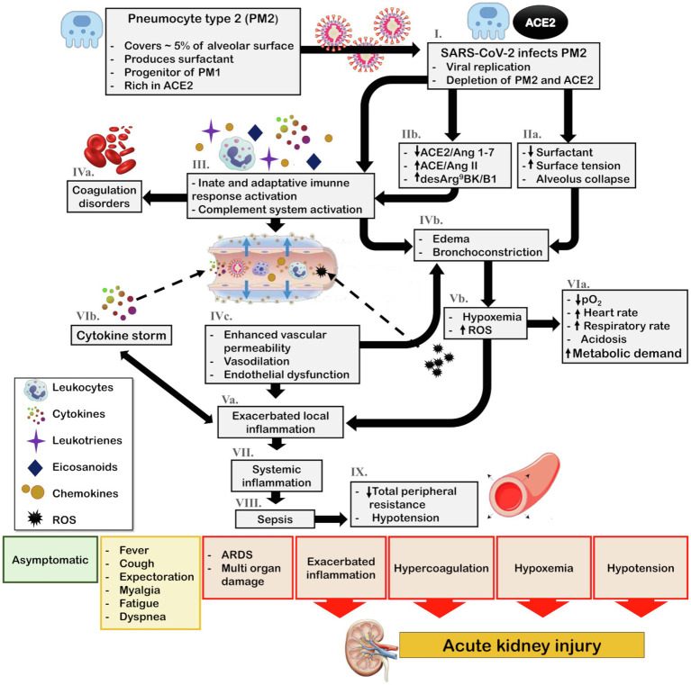
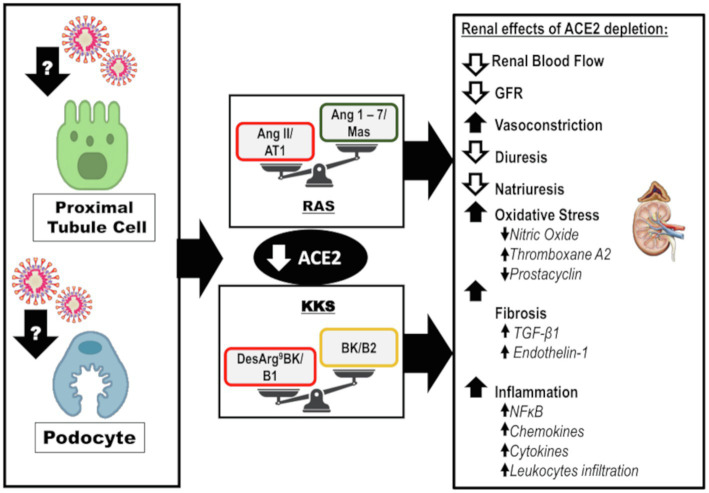
Coronavirus disease 2019 (COVID-19) was first reported in late December 2019 in Wuhan, China. The etiological agent of this disease is severe acute respiratory syndrome coronavirus 2 (SARS-CoV-2), and the high transmissibility of the virus led to its rapid global spread and a major pandemic (ongoing at the time of writing this review). The clinical manifestations of COVID-19 can vary widely from non-evident or minor symptoms to severe acute respiratory syndrome and multi-organ damage, causing death. Acute kidney injury (AKI) has been recognized as a common complication of COVID-19 and in many cases, kidney replacement therapy (KRT) is required. The presence of kidney abnormalities on hospital admission and the development of AKI are related to a more severe presentation of COVID-19 with higher mortality rate. The high transmissibility and the broad spectrum of clinical manifestations of COVID-19 are in part due to the high affinity of SARS-CoV-2 for its receptor, angiotensin (Ang)-converting enzyme 2 (ACE2), which is widely expressed in human organs and is especially abundant in the kidneys. A debate on the role of ACE2 in the infectivity and pathogenesis of COVID-19 has emerged: Does the high expression of ACE2 promotes higher infectivity and more severe clinical manifestations or does the interaction of SARS-CoV-2 with ACE2 reduce the bioavailability of the enzyme, depleting its biological activity, which is closely related to two important physiological systems, the renin-angiotensin system (RAS) and the kallikrein-kinin system (KKS), thereby further contributing to pathogenesis. In this review, we discuss the dual role of ACE2 in the infectivity and pathogenesis of COVID-19, highlighting the effects of COVID-19-induced ACE2 depletion in the renal physiology and how it may lead to kidney injury. The ACE2 downstream regulation of KKS, that usually receives less attention, is discussed. Also, a detailed discussion on how the triad of symptoms (respiratory, inflammatory, and coagulation symptoms) of COVID-19 can indirectly promote renal injury is primary aborded.

Keywords: acute kidney injury; angiotensin-converting enzyme 2; coronavirus disease 2019; kallikrein-kinin system; renin-angiotensin system; severe acute respiratory syndrome coronavirus 2.


Figures

Figure 1
Figure 1
Angiotensin-converting enzyme 2 has a catalytic role in RAS and KKS. (A) Renin converts the precursor, angiotensinogen, into angiotensin I. In a classic pathway, angiotensin I is cleaved by ACE to form Angiotensin II. ACE2 can biosynthesize angiotensin 1–7 by two distinct pathways: acting directly on angiotensin II or alternatively converting angiotensin I into angiotensin 1–9 that is further cleaved by ACE, generating angiotensin 1–7. (B) The precursor kininogen is cleaved by kallikrein to form the active peptide, bradykinin that is rapidly degraded by ACE, or in an alternative pathway, can be converted to desArg9bradykinin by CPM and CPN. ACE2 can inactivate desArg9bradykinin. ACE, angiotensin-converting enzyme; ACE2, angiotensin-converting enzyme 2; CPM, carboxypeptidase M; CPN, carboxypeptidase N; KKS, kallikrein-kinin system; and RAS, renin-angiotensin system.
Figure 2
Figure 2
Structure of SARS-CoV-2 and the proposed mechanisms of cell entry and replication. (A) SARS-CoV-2 is composed of RNA contained in nucleocapsids; the envelope retains the genetic material and contains the membrane protein and hemagglutinin esterase dimers. Attached to the envelope is the spike protein (S) responsible for receptor binding. (B) Enzymes reported to participate in the cell entry mechanism of SARS-CoV-2. (C) (I.) SARS-CoV-2 recognizes its receptor, ACE2. The short pathway is possible if S had been primed by furin during biosynthesis and in the presence of TMPRSS2 and/trypsin that cleave S1; (II.) the virus fuses with plasmatic membrane; (III.) releasing its genetic material into the cytosol; (IV.) RNA transcription and replication occur in the cytosol, while the structural proteins are biosynthesized in the endoplasmic reticulum and Golgi apparatus, at this point, furin can prime S at S1/S2; and (V.) New genetic material is encapsulated by envelope and structural proteins generating new virions (VI.) that will be released from the host cell. (D) (I.) The SARS-CoV-2 recognizes its receptor, ACE2. If S had not been primed, a second pathway is activated and (II.) the virus is endocytosed; (III.) Owing to the decreased pH, cathepsin L can be activated and cleaves S1, promoting fusion of the SARS-CoV-2 with the endosome membrane and (IV.) the release of viral genetic material; (V.) RNA transcription and replication occur in the cytosol while biosynthesis of the structural proteins occurs in the endoplasmic reticulum and Golgi apparatus. In this representation, there is no furin to prime S at S1/S2; and (VI.) The genetic material is encapsulated by envelope and structural proteins, generating new virions (VII.) that will be released from the host cell. ACE2, angiotensin-converting enzyme 2; S, spike protein; SARS-CoV-2, severe acute respiratory syndrome coronavirus 2; and TMPRSS2, transmembrane serine protease 2.
Figure 3
Figure 3
Schematic representation of COVID-19 pathophysiology and its indirect effects on kidney. The main target of SARS-CoV-2 in the lungs is PM2. (I.) Viral infection and replication can culminate the depletion of PM2 and ACE2 reservoir. Consequently, (IIa.) surfactant production diminishes, leading to an increase in surface tension and alveolar collapse. Additionally, (IIb.) SARS-CoV-2 infection associated with ACE2/Ang 1–7 downregulation and ACE/Ang II and desArg9BK/B1 exacerbation promotes the (III.) activation of the innate and adaptative immune response and complement system leading to recruitment of leukocytes and release of cytokines, chemokines, eicosanoids, and leukotrienes. The complement system and eicosanoids promote (IVa.) coagulation disorders and the leukotrienes in association with increased surface tension contribute to (IVb.) bronchoconstriction. Moreover, the local inflammation culminates in (IVc.) increased vascular permeability, vasodilation, and endothelial dysfunction, thereby enhancing leukocyte recruitment, and leading to (Va.) exacerbated local inflammation what also contribute to (IVb.) bronchoconstriction and edema. Furthermore, bronchoconstriction and edema lead to (Vb.) hypoxia and ROS generation, which contributes to (VIa.) cardiorespiratory alterations that increase metabolic demand. Most importantly, ROS generation feeds the cycle of (Va.) enhanced inflammation. This inflammatory state leads to the (VIb.) cytokine storm and due to (IVc.) enhanced vascular permeability, viral particles, leukocytes, ROS, and cytokines can reach the blood stream, ultimately causing (VII.) systemic inflammation, (VIII.) sepsis, and consequently (IX.) hypotension. Depending on the patient health status, the steps in the pathophysiological cascade that are activated, and the intensity of the immune response, the clinical manifestations can vary from asymptomatic and mild symptoms, such as fever, cough, and myalgia, to severe symptoms, including acute respiratory distress and multi-organ damage. In this scenario, exacerbated inflammation, coagulation disorders, hypoxemia, and hypotension contribute to acute kidney injury (AKI). ACE, angiotensin-converting enzyme; ACE2, angiotensin-converting enzyme 2; Ang 1–7, angiotensin 1–7; Ang II, angiotensin II; B1, kinin receptor type 1; COVID-19, coronavirus disease 2019; desArg9BK, desArg9bradykinin; PM1, pneumocytes type I; PM2, pneumocytes type II; ROS, reactive oxygen species; and SARS-CoV-2, severe acute respiratory syndrome coronavirus 2.
Figure 4
Figure 4
Direct effects of SARS-CoV-2 on kidneys: Infection and disruption of the downstream mechanisms regulated by ACE2. It is likely that SARS-CoV-2 infects the kidney by directly targeting the proximal tubule cells and podocytes. The infection can lead to depletion of ACE2 and its biological functions at the intrarenal level, which may lead to exacerbated actions of the ACE/Ang II/AT1 axis of RAS and desArg9BK/B1 axis of KKS. This impairment leads to reduced renal blood flow, GFR, diuresis, and natriuresis. However, there is an increase in vasoconstriction. The oxidative stress is enhanced due to a decrease in NO levels, interfering in the balance between prostacyclin and thromboxane A2. Furthermore, fibrosis is enhanced in response to TGF-β1 and endothelin-1. Finally, inflammation is upregulated along with augmented levels of chemokines, cytokines, and leukocyte recruitment. ACE2, angiotensin-converting enzyme 2; AT1, angiotensin II receptor type 1; BK, bradykinin; desArg9BK, desArg9bradykinin; B1, kinin receptor type 1; B2, kinin receptor type 2; GFR, glomerular filtration rate; KKS, kallikrein-kinin system; and RAS, renin-angiotensin system.

Similar articles

Cited by

References

    1. Akcay A., Nguyen Q., Edelstein C. L. (2009). Mediators of inflammation in acute kidney injury. Mediators Inflamm. 2009:137072. 10.1155/2009/137072, PMID: - DOI - PMC - PubMed
    1. Aragão D. S., Cunha T. S., Arita D. Y., Andrade M. C., Fernandes A. B., Watanabe I. K., et al. . (2011). Purification and characterization of angiotensin converting enzyme 2 (ACE2) from murine model of mesangial cell in culture. Int. J. Biol. Macromol. 49, 79–84. 10.1016/j.ijbiomac.2011.03.018, PMID: - DOI - PMC - PubMed
    1. Batlle D., Soler M. J., Sparks M. A., Hiremath S., South A. M., Welling P. A., et al. . (2020). Acute kidney injury in COVID-19: emerging evidence of a distinct pathophysiology. J. Am. Soc. Nephrol. 31, 1380–1383. 10.1681/ASN.2020040419, PMID: - DOI - PMC - PubMed
    1. Benedetti C., Waldman M., Zaza G., Riella L. V., Cravedi P. (2020). COVID-19 and the kidneys: an update. Front. Med. 7:423. 10.3389/fmed.2020.00423, PMID: - DOI - PMC - PubMed
    1. Bhatraju P. K., Ghassemieh B. J., Nichols M., Kim R., Jerome K. R., Nalla A. K., et al. . (2020). Covid-19 in critically ill patients in the Seattle region – case series. N. Engl. J. Med. 382, 2012–2022. 10.1056/NEJMoa2004500, PMID: - DOI - PMC - PubMed