. 2021 Aug 21:2021:8231559.
doi: 10.1155/2021/8231559. eCollection 2021.

Cholecystokinin Expression in the Development of Myocardial Hypertrophy

Affiliations

Cholecystokinin Expression in the Development of Myocardial Hypertrophy

Zhongshu Han et al. Scanning. .

Abstract

Background: Expression of cholecystokinin is found in myocardial tissues as a gastrointestinal hormone and may be involved in cardiovascular regulation. However, it is unclear whether there is an increase in cholecystokinin expression in myocardial hypertrophy progression induced by abdominal aortic constriction. The study is aimed at exploring the relationship between cholecystokinin expression and myocardial hypertrophy.

Methods: We randomly divided the 70 Sprague-Dawley rats into two groups: the sham operation group and the abdominal aortic constriction group. The hearts of rats were measured by echocardiography, and myocardial tissues and blood were collected at 4 weeks, 8 weeks, and 12 weeks after surgery. Morphological changes were assessed by microscopy. The cholecystokinin expression was evaluated by immunochemistry, Western blotting, quantitative real-time polymerase chain reaction, and enzyme-linked immunosorbent assay.

Results: The relative protein levels of cholecystokinin were significantly increased in the abdominal aortic constriction groups compared with the corresponding sham operation groups at 8 weeks and 12 weeks. The cholecystokinin mRNA in the abdominal aortic constriction groups was significantly higher than the time-matched sham operation groups. Changes in the left ventricular wall thickness were positively correlated with the relative protein levels of cholecystokinin and the mRNA of cholecystokinin.

Conclusions: The development of myocardial hypertrophy can affect the cholecystokinin expression of myocardial tissues.


Conflict of interest statement

The authors declare that they have no competing interests.

Figures
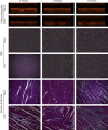
Figure 1
Figure 1
Echocardiography and pathological examination of rat heart. Echocardiography (first and second panels), H&E (third and fourth panels), and Masson's trichrome (fifth and sixth panels) staining of myocardial tissues at 4, 8, and 12 weeks after surgery. Sham: sham operation group; AAC: abdominal aortic constriction; H&E: haematoxylin and eosin. Pathological images were photographed at ×200 magnification.
Figure 2
Figure 2
CCK expression levels were detected by immunohistochemical staining (×400 magnification). Sham: sham operation group; AAC: abdominal aortic constriction.
Figure 3
Figure 3
The myocardial tissues of the left ventricular were collected and analyzed by RT-PCR and Western blotting; the plasma samples were collected at the end of experiment and analyzed by ELISA. (a) Analysis of CCK and GAPDH protein expression from myocardial tissues of the left ventricular by Western blotting. (b) Bar graph shows levels of CCK mRNA in myocardial tissue. (c) Bar graph shows relative intensity of CCK to GAPDH in myocardial tissue. (d) The bars indicate the plasma CCK levels. CCK: cholecystokinin; GAPDH: glyceraldehyde-3-phosphate dehydrogenase; ELISA: enzyme-linked immunosorbent assay; sham: sham operation group; AAC: abdominal aortic constriction. P < 0.05, ∗∗P < 0.01, compared with sham groups at the same time point; #P < 0.01, versus the 8 weeks and 12 weeks. AAC groups; ##P < 0.01, versus the 4 weeks and 12 weeks AAC groups; ###P < 0.01, versus the 4 weeks and 8 weeks AAC groups; &P < 0.01, versus the 8 weeks and 12 weeks AAC groups.
Figure 4
Figure 4
Associations between expression of IVSd and C-CK in model rat. (a) Correlation between IVSd and CCK mRNA (r = 0.694, P < 0.001); (b) correlation between IVSd and relative protein level of CCK (r = 0.826, P < 0.001). CCK: cholecystokinin; IVSd: end-diastolic interventricular septal thickness.
Figure 5
Figure 5
Associations between expression of LVPWd and CCK in model rat. (a) Correlation between LVPWd and CCK mRNA (r = 0.646, P < 0.001); (b) Correlation between LVPWd and relative protein level of CCK (r = 0.692, P < 0.001). CCK: cholecystokinin; LVPWd: left ventricular end-diastolic posterior wall thickness.

Similar articles

Cited by

References

    1. Goetze J. P. B-Type natriuretic peptide: from posttranslational processing to clinical measurement. Clinical Chemistry. 2012;58(1):83–91. doi: 10.1373/clinchem.2011.165696. - DOI - PubMed
    1. Deftos L. J., Burton D. W., Brandt D. W. Parathyroid hormone-like protein is a secretory product of atrial myocytes. The Journal of Clinical Investigation. 1993;92(2):727–735. doi: 10.1172/JCI116643. - DOI - PMC - PubMed
    1. Ito H., Hirata Y., Adachi S., et al. Endothelin-1 is an autocrine/paracrine factor in the mechanism of angiotensin II-induced hypertrophy in cultured rat cardiomyocytes. The Journal of Clinical Investigation. 1993;92(1):398–403. doi: 10.1172/JCI116579. - DOI - PMC - PubMed
    1. Dschietzig T., Richter C., Bartsch C., et al. The pregnancy hormone relaxin is a player in human heart failure. The FASEB Journal. 2001;15(12):2187–2195. doi: 10.1096/fj.01-0070com. - DOI - PubMed
    1. Wang C., Dong X. Y., Wei L. M., et al. The relationship of Appetite-Regulating hormones in the development of cardiac cachexia. International Heart Journal. 2019;60(2):384–391. doi: 10.1536/ihj.18-131. - DOI - PubMed

Substances

LinkOut - more resources