

Turning the Actin Nucleating Compound Miuraenamide into Nucleation Inhibitors
- PMID: 34497907
- PMCID: PMC8412923
- DOI: 10.1021/acsomega.1c02838
Turning the Actin Nucleating Compound Miuraenamide into Nucleation Inhibitors
Abstract
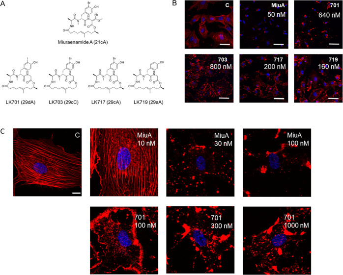
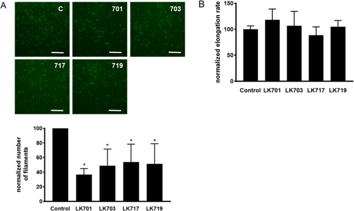
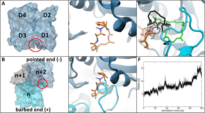
Natural compounds that either increase or decrease polymerization of actin into filaments have become indispensable tools for cell biology. However, to date, it was not possible to use them as therapeutics due to their overall cytotoxicity and their unfavorable pharmacokinetics. Furthermore, their synthesis is in general quite complicated. In an attempt to find simplified analogues of miuraenamide, an actin nucleating compound, we identified derivatives with a paradoxical inversion of the mode of action: instead of increased nucleation, they caused an inhibition. Using an extensive computational approach, we propose a binding mode and a mode of action for one of these derivatives. Based on our findings, it becomes feasible to tune actin-binding compounds to one or the other direction and to generate new synthetic actin binders with increased functional selectivity.
© 2021 The Authors. Published by American Chemical Society.
Conflict of interest statement
The authors declare no competing financial interest.
Figures
Similar articles
-
Actin stabilization in cell migration.Front Cell Dev Biol. 2022 Aug 11;10:931880. doi: 10.3389/fcell.2022.931880. eCollection 2022. Front Cell Dev Biol. 2022. PMID: 36035985 Free PMC article.
-
Actin stabilizing compounds show specific biological effects due to their binding mode.Sci Rep. 2019 Jul 5;9(1):9731. doi: 10.1038/s41598-019-46282-w. Sci Rep. 2019. PMID: 31278311 Free PMC article.
-
Persistent inhibition of pore-based cell migration by sub-toxic doses of miuraenamide, an actin filament stabilizer.Sci Rep. 2017 Nov 27;7(1):16407. doi: 10.1038/s41598-017-16759-7. Sci Rep. 2017. PMID: 29180826 Free PMC article.
-
Cellular control of actin nucleation.Annu Rev Cell Dev Biol. 2002;18:247-88. doi: 10.1146/annurev.cellbio.18.040202.112133. Epub 2002 Apr 2. Annu Rev Cell Dev Biol. 2002. PMID: 12142287 Review.
-
Microfilament dynamics: regulation of actin polymerization by actin-fragmin kinase and phosphatases.Adv Enzyme Regul. 1995;35:199-227. doi: 10.1016/0065-2571(94)00013-s. Adv Enzyme Regul. 1995. PMID: 7572344 Review.
Cited by
-
Cytoskeletal dysregulation and neurodegenerative disease: Formation, monitoring, and inhibition of cofilin-actin rods.Front Cell Neurosci. 2022 Sep 22;16:982074. doi: 10.3389/fncel.2022.982074. eCollection 2022. Front Cell Neurosci. 2022. PMID: 36212686 Free PMC article. Review.
-
Actin stabilization in cell migration.Front Cell Dev Biol. 2022 Aug 11;10:931880. doi: 10.3389/fcell.2022.931880. eCollection 2022. Front Cell Dev Biol. 2022. PMID: 36035985 Free PMC article.
-
Genomic Analysis of the Rare Slightly Halophilic Myxobacterium "Paraliomyxa miuraensis" SMH-27-4, the Producer of the Antibiotic Miuraenamide A.Microorganisms. 2023 Feb 1;11(2):371. doi: 10.3390/microorganisms11020371. Microorganisms. 2023. PMID: 36838335 Free PMC article.
-
Syntheses of Marine Natural Products via Matteson Homologations and Related Processes.Mar Drugs. 2025 Jan 2;23(1):20. doi: 10.3390/md23010020. Mar Drugs. 2025. PMID: 39852522 Free PMC article. Review.
-
A Novel Interaction of Slug (SNAI2) and Nuclear Actin.Cells. 2024 Apr 17;13(8):696. doi: 10.3390/cells13080696. Cells. 2024. PMID: 38667311 Free PMC article.
References
LinkOut - more resources
Full Text Sources
