. 2021 Sep;70(9):001413.
doi: 10.1099/jmm.0.001413.

Challenges on the development of a pseudotyping assay for Zika glycoproteins

Affiliations

Challenges on the development of a pseudotyping assay for Zika glycoproteins

Fernando Ruiz-Jiménez et al. J Med Microbiol. 2021 Sep.

Abstract

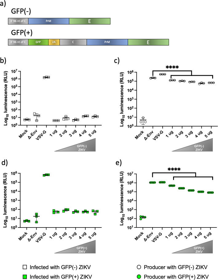
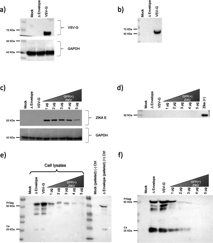
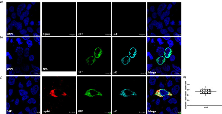
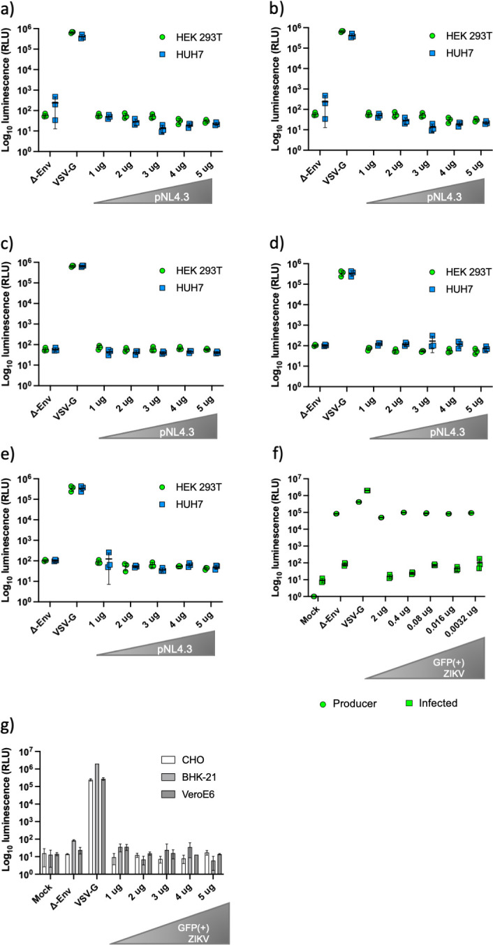
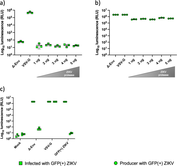
Introduction. Zika virus (ZIKV) emerged as a public health concern on the American continent during late 2015. As the number of infected grew so did the concerns about its capability to cause long-term damage especially with the appearance of the congenital Zika syndrome (CZS). Proteins from the TAM family of receptor tyrosine kinases (RTKs) were proposed as the cellular receptors, however, due to the ability of the virus to infect a variety of cell lines different strategies to elucidate the tropism of the virus should be investigated.Hypothesis. Pseudotyping is a powerful tool to interrogate the ability of the glycoprotein (GP) to permit entry of viruses.Aim. We aimed to establish a highly tractable pseudotype model using lenti- and retro-viral backbones to investigate the entry pathway of ZIKV.Methodology. We used different glycoprotein constructs and different lenti- or retro-viral backbones, in a matrix of ratios to investigate production of proteins and functional pseudotypes.Results. Varying the ratio of backbone and glycoprotein plasmids did not yield infectious pseudotypes. Moreover, the supplementation of the ZIKV protease or the substitution of the backbone had no positive impact on the infectivity. We showed production of the proteins in producer cells implying the lack of infectious pseudotypes is due to a lack of successful glycoprotein incorporation, rather than lack of protein production.Conclusion. In line with other reports, we were unable to successfully produce infectious pseudotypes using the variety of methods described. Other strategies may be more suitable in the development of an efficient pseudotype model for ZIKV and other flaviviruses.

Keywords: Zika virus; glycoprotein; pseudotype.


Conflict of interest statement

The authors declare that there are no conflicts of interest.

Figures

Fig. 1.
Fig. 1.
Addition of GFP into the ZIKV GP construct had no effect on measured luciferase activity in both producer and infected cells. (a) Schematic of the two ZIKV glycoprotein constructs tested. Luciferase activity measured in (b) infected and (c) producer cells using the GFP(-) ZIKV construct. Luciferase activity measured in (d) infected and (e) producer cells using the GFP(+) ZIKV construct. VSV-G and Δ envelope were used as positive and negative controls, respectively. Graphs show mean±sd of three independent experiments.
Fig. 2.
Fig. 2.
Detection of viral proteins in cell lysates and pelleted pseudotypes by Western blot. The presence of the positive control VSV-G protein was visualized in (a) cell lysates and (b) pelleted pseudotypes. Intensity of ZIKV envelope staining in cell lysates (c) was inversely related to the amount of plasmid transfected. No ZIKV envelope was visible in pelleted pseudotypes (d). Intensity of HIV capsid precursor protein (p55) in cell lysates (e) was inversely related to the amount of GFP(+) ZIKV plasmid transfected. Low levels of processed p24 capsid protein were detected in the pelleted pseudotypes (f).
Fig. 3.
Fig. 3.
Immunofluorescence of transfected HEK 293 T cells against HIV-1 and ZIKV proteins. (a) HEK 293T control cells showing no cross reactivity or background fluorescence. (b) HEK 293 T cells transfected with the GFP(+) ZIKV glycoprotein plasmid expressed both the reporter protein (GFP, green) and the viral GP/ E (α-E, cyan). (c) HEK 293 T cells transfected with both GFP(+) ZIKV glycoprotein plasmid and pNL4.3 HIV-1 plasmid expressed ZIKV E, and the capsid p24 HIV-1 protein (α-p24, red). (d) Pearson's correlation coefficient graph showing a moderate colocalization between ZIKV E and HIV-1 p24 (r=0.53). Scale bar of 10 µm.
Fig. 4.
Fig. 4.
Different cell lines treated with ZIKV pseudotypes. HEK 293T and HUH7 cells infected with pseudotypes produced with different ratios of GFP(+) ZIKV glycoprotein and pNL4.3. (a) 1 µg of GFP(+) ZIKV with a range (1–5 µg) of pNL4.3. (b) 2 µg of GFP(+) ZIKV with a range (1–5 µg) of pNL4.3. (c) 3 µg of GFP(+) ZIKV with a range (1–5 µg) of pNL4.3. (d) 4 µg of GFP(+) ZIKV with a range (1–5 µg) of pNL4.3. (e) 5 µg of GFP(+) ZIKV with a range (1–5 µg) of pNL4.3. Pseudotypes were not infectious in either cell line, regardless of amount of plasmid used. (f) HEK 293 T cells were transfected with a fixed pNL4.3 plasmid amount (2 µg) and fivefold dilutions of the ZIKV GP plasmid (2, 0.4, 0.08, 0.016 and 0.0032 µg). No infectious ZIKV pseudotypes were generated. (g) Mammalian cell lines CHO, BHK-21 and VeroE6 were treated with pseudotypes produced using increasing concentrations of GFP(+) ZIKV plasmid (1–5 µg) in HEK 293 T cells. Again, no infectivity was measured. VSV-G and Δ envelope were used as positive and negative controls, respectively. Graphs show mean±sd of three independent experiments.
Fig. 5.
Fig. 5.
Luciferase activity measured in the producer and infected cells supplemented with viral protease plasmid. HEK 293 T cells were co-transfected with 2 µg of pNL4.3 and 2 µg of GFP(+) ZIKV. Increasing amounts (1–5 µg) of the NS2B/NS3 protease plasmid were added. Luciferase activity measured in (a) infected and (b) producer cells showed no infectious pseudotypes produced and no reduction in luciferase activity in the producer cells. (c) Murine Leukaemia Virus (MLV) backbone was tested at 2 µg with 2 µg GFP(+) ZIKV to identify if the backbone had an effect on infectivity. No infectious particles were produced. VSV-G and Δ envelope were used as positive and negative controls, respectively. Graphs show mean±sd of three independent experiments.

References

    1. Wang A, Thurmond S, Islas L, Hui K, Hai R. Zika virus genome biology and molecular pathogenesis. Emerg Microbes Infect. 2017;6:e13. - PMC - PubMed
    1. Heinz F, Stiasny K. The antigenic structure of Zika virus and its relation to other Flaviviruses: implications for infection and immunoprophylaxis. Microbiol Mol Biol Rev. 2017;81:e00055-16. doi: 10.1128/MMBR.00055-16. - DOI - PMC - PubMed
    1. Imperato PJ. The Convergence of a virus, mosquitoes, and human travel in globalizing the Zika epidemic. J Community Health. 2016;41:674–679. doi: 10.1007/s10900-016-0177-7. - DOI - PubMed
    1. MacNamara FN. Zika virus: A report on three cases of human infection during an epidemic of jaundice in Nigeria. Trans R Soc Trop Med Hyg. 1954;48:139–145. doi: 10.1016/0035-9203(54)90006-1. - DOI - PubMed
    1. Gebre Y, Forbes N, Gebre T. Zika virus infection, transmission, associated neurological disorders and birth abnormalities: A review of progress in research, priorities and knowledge gaps. Asian Pac J Trop Biomed. 2016;6:815–824. doi: 10.1016/j.apjtb.2016.08.008. - DOI

Publication types