Review
. 2022 Jan 1;136(1):181-205.
doi: 10.1097/ALN.0000000000003943.

Perioperative Pulmonary Atelectasis: Part I. Biology and Mechanisms

Affiliations
Review

Perioperative Pulmonary Atelectasis: Part I. Biology and Mechanisms

Congli Zeng et al. Anesthesiology. .

Abstract

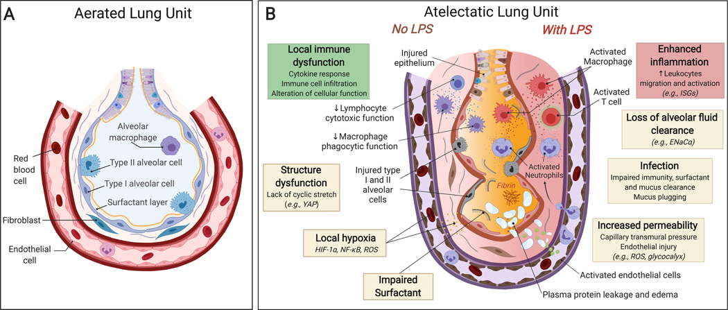
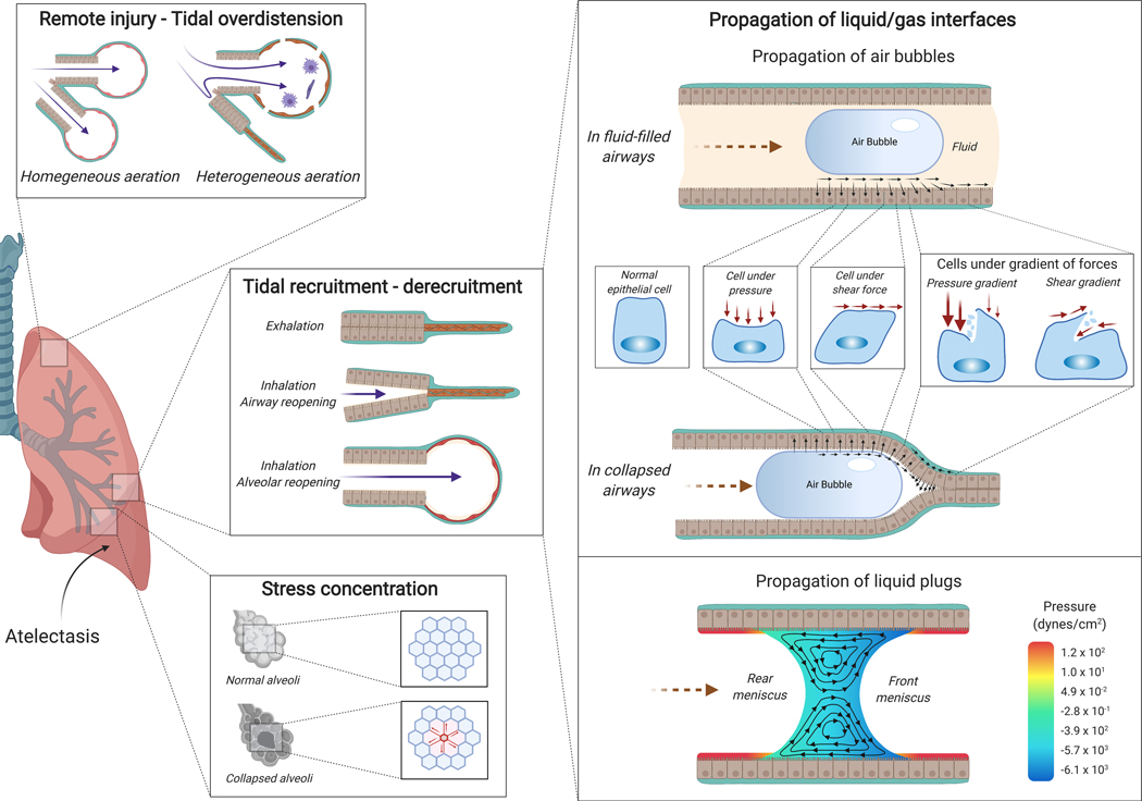
Pulmonary atelectasis is common in the perioperative period. Physiologically, it is produced when collapsing forces derived from positive pleural pressure and surface tension overcome expanding forces from alveolar pressure and parenchymal tethering. Atelectasis impairs blood oxygenation and reduces lung compliance. It is increasingly recognized that it can also induce local tissue biologic responses, such as inflammation, local immune dysfunction, and damage of the alveolar-capillary barrier, with potential loss of lung fluid clearance, increased lung protein permeability, and susceptibility to infection, factors that can initiate or exaggerate lung injury. Mechanical ventilation of a heterogeneously aerated lung (e.g., in the presence of atelectatic lung tissue) involves biomechanical processes that may precipitate further lung damage: concentration of mechanical forces, propagation of gas-liquid interfaces, and remote overdistension. Knowledge of such pathophysiologic mechanisms of atelectasis and their consequences in the healthy and diseased lung should guide optimal clinical management.


Conflict of interest statement

Conflicts of Interest: none.

Figures

Fig. 1:
Fig. 1:. Pressures and forces acting on alveolar and bronchiolar walls and visceral pleura surface.
Fi = inward tethering; Fo = outward tethering; Fw = circumferential component of force applied by the layer of surface-active fluid; Palv = alveolar pressure; Pi = inside pressure; Po = outside pressure; Ppl = pleural pressure.
Fig. 2:
Fig. 2:. Mechanisms producing atelectasis in the perioperative period.
(A) Normal lung unit in awake conditions: adequate inspiratory (Pi) and expiratory (Pe) intraluminal pressure and bronchiolar or alveolar tethering stress associated with negative pleural pressure (Ppl) allow for the normal opening of the bronchiole and a normal alveolar ventilation (⩒A). Alveolar gas absorption is physiological due to physiological ⩒A/Q̇ and atmospheric FIO2. Normal surfactant reduces alveolar surface tension. (B) Lung unit exposed to perioperative atelectasis: increase in pleural pressure (Ppl) due to extrinsic or intrinsic compression (formula image
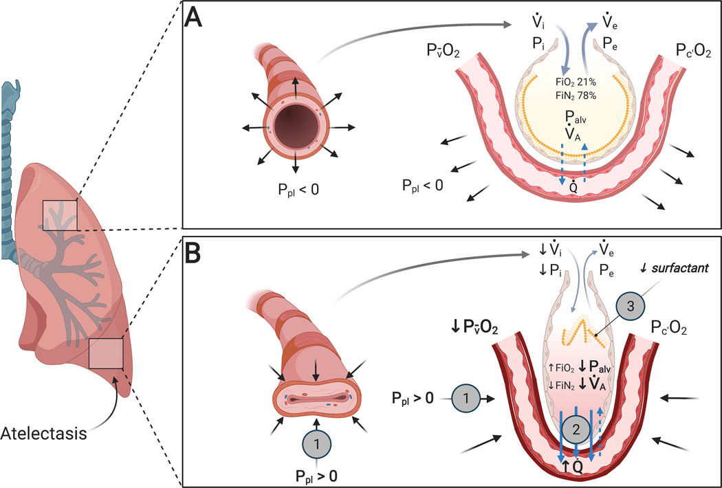
) is responsible for loss of expansion and reduced alveolar ventilation (⩒A). Increased alveolar gas absorption (formula image) reduces intraluminal alveolar pressure (Palv). Low ⩒A/Q̇, high FIO2 and low mixed venous oxygen partial pressure (P v¯O2) may participate in such gas exchange imbalance. Quantitative or qualitative surfactant impairment leads to higher surface tension and facilitates alveolar collapse (formula image). Pc’O2 = end-capillary oxygen partial pressure.
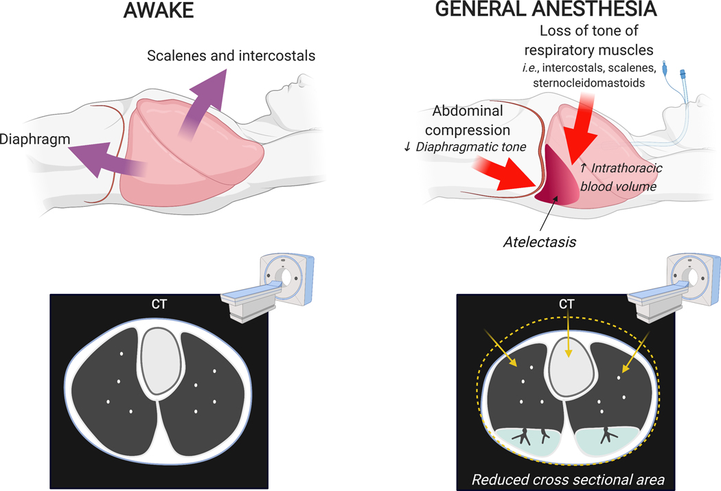
Fig. 3:
Fig. 3:. Changes in chest wall shape due to general anesthesia in a supine patient.
During awake spontaneous breathing, contraction of diaphragm and accessory muscles of respiration maintain lung expansion. Loss of muscular tone during anesthesia is associated with cephalad motion of the dependent diaphragm, reduction in cross-sectional chest area, and generation of non-gravitational compressive forces (i.e., cephalocaudal gradients). Together with gravitational forces and potential increase in intrathoracic blood volume, these factors contribute to reduction of lung volume and lung collapse particularly on the dorsal and basal lung regions.
Fig. 4:
Fig. 4:. Pulmonary pressure-volume curve during inhalation and exhalation showing lung hysteresis.
The shape of the lung pressure-volume curve is sigmoidal. There are three main portions of the curve. The initial portion (blue) of lung recruitment at low pressures and volumes is related to low compliance (i.e., the change in volume divided by the change in pressure is low). This is followed by a portion with linear relationship between volume and pressure (ochre) with higher compliance. Finally, hyperinflation ensues at high pressures and volumes with return of lower compliance (red). The transition between the first and second portions indicate that critical opening pressures for a large number of bronchoalveolar regions has been reached (lower inflection point). Note the higher pressure to reach the same lung volume during inhalation than exhalation. Modifed from Radford EP Jr.
Fig. 5:
Fig. 5:. Pressure-volume relationship for the chest wall, lungs and their combination (respiratory system) in normal (A) and obese (B) subjects in supine positions.
(A) The shape of the respiratory system pressure-volume curve reflects the balance of the forces from the chest wall and lung parenchyma. In normals, functional residual capacity is reduced as compared to the upright position but large enough to locate an operating range of the respiratory system pressure-volume curve within a region of high compliance (dashed yellow rectangle). (B) In obese subjects, increased weight of the chest wall and abdomen shift the chest wall pressure-volume curve to the right at similar chest wall compliances. Combined with the substantial reduction of functional residual capacity (FRC), the operating range of the respiratory system pressure-volume curve moves to a region of low compliance (dashed yellow rectangle). This occurs even in the presence of the same normal pressure-volume curve of the lungs. Modifed from Behazin et al.
Fig. 6:
Fig. 6:. Atelectasis-associated regional biological injury.
(A) Normally aerated lung. (B) Atelectatic Lung. There is local immune dysfunction with dysregulated cytokine secretion and impaired function of immune cell and surfactant, in part associated with local hypoxia and lack of cyclic stretch. Endotoxemia (=systemic lipopolysaccharide, LPS) enhances inflammatory responses characterized by marked immune cell infiltration and activation. In addition, atelectasis leads to structure dysfunction accompanied by loss of alveolar fluid clearance and increased protein permeability, flooding of the airspace with protein-rich pulmonary edema fluid, and potentially increased susceptibility to infection.
Fig. 7:
Fig. 7:. Atelectasis-associated regional mechanical injury.
Airway or alveolar injury can occur during tidal recruitment-derecruitment. Propagation of gas-liquid interfaces is a potential mechanism. During inhalation, air propagates into a fluid-filled airway (right upper panel) or a collapsed airway (right middle panel) generating mechanical forces at the interface of air bubble and airway with resulting cell deformations due to normal pressure, shear stress, and their gradients. The pressure gradient is likely the major determinant of injury. Propagation of liquid plugs and rupture of the liquid menisci also generate abnormally large mechanical forces in the area of smallest film thickness where the front meniscus converges to a precursor film (right lower panel). Stress concentration (left bottom panel) is another potential mechanism for atelectasis-related lung injury. In normally expanded lungs, the alveoli are ventilated homogeneously. During atelectasis, however, collapsed areas may result in stress concentration, locally multiplying the stress around the atelectatic regions. Atelectasis also leads to the redistribution of tidal volume from atelectatic to aerated areas resulting in remote tidal overdistension (left upper panel).

Comment in

  • Perioperative Pulmonary Atelectasis: Comment.
    van Egmond J, Speight C, Roberts JHM, Patel A, van Rijn CM, Coulthard M. van Egmond J, et al. Anesthesiology. 2022 Jul 1;137(1):125-126. doi: 10.1097/ALN.0000000000004231. Anesthesiology. 2022. PMID: 35486841 No abstract available.

References

    1. Bendixen HH, Hedley-Whyte J, Laver MB: Impaired oxygenation in surgical patients during general anesthesia with controlled ventilation. A concept of atelectasis. N Engl J Med 1963; 269:991–6 - PubMed
    1. Brismar B, Hedenstierna G, Lundquist H, Strandberg A, Svensson L, Tokics L: Pulmonary densities during anesthesia with muscular relaxation--a proposal of atelectasis. Anesthesiology 1985; 62:422–8 - PubMed
    1. Futier E, Constantin J-M, Paugam-Burtz C, Pascal J, Eurin M, Neuschwander A, Marret E, Beaussier M, Gutton C, Lefrant J-Y, Allaouchiche B, Verzilli D, Leone M, De Jong A, Bazin J-E, Pereira B, Jaber S: A Trial of Intraoperative Low-Tidal-Volume Ventilation in Abdominal Surgery. N Engl J Med 2013; 369:428–37 - PubMed
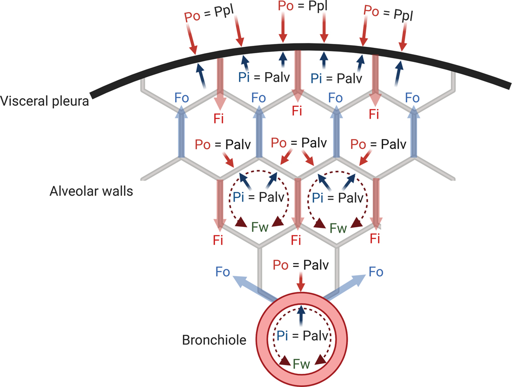
    1. Mead J, Takishima T, Leith D: Stress distribution in lungs: a model of pulmonary elasticity. J Appl Physiol 1970; 28:596–608 - PubMed
    1. Burgstaller G, Oehrle B, Gerckens M, White ES, Schiller HB, Eickelberg O: The instructive extracellular matrix of the lung: basic composition and alterations in chronic lung disease. Eur Respir J 2017; 50 - PubMed

Publication types

MeSH terms