. 2022 Mar 29;145(1):64-75.
doi: 10.1093/brain/awab311.

Emerging roles for dynamic aquaporin-4 subcellular relocalization in CNS water homeostasis

Affiliations

Emerging roles for dynamic aquaporin-4 subcellular relocalization in CNS water homeostasis

Mootaz M Salman et al. Brain. .

Abstract

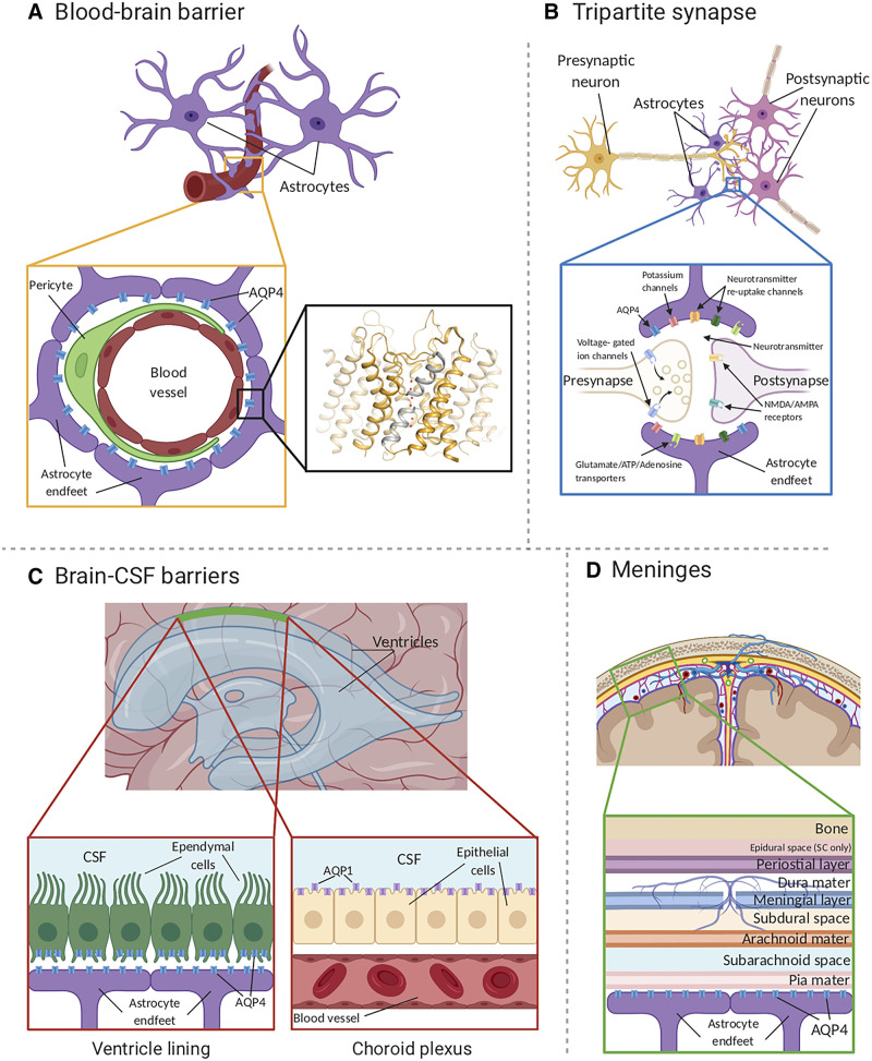
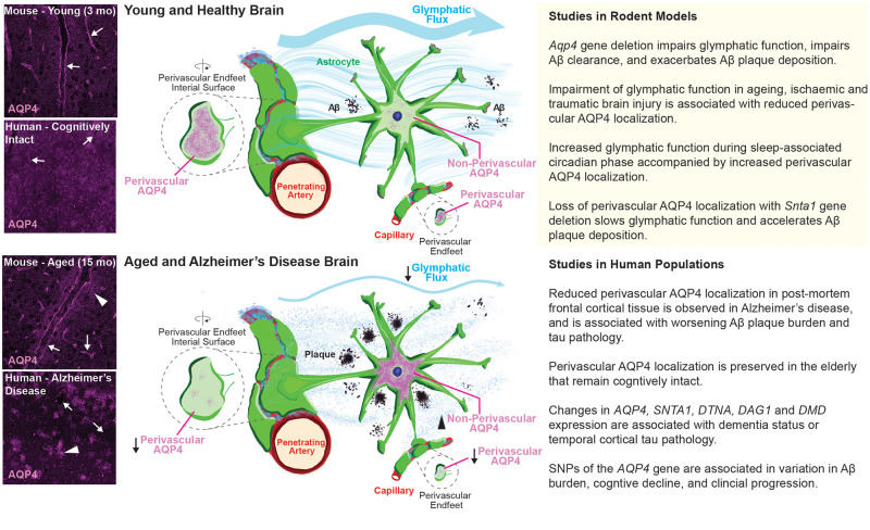
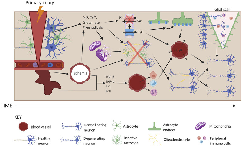
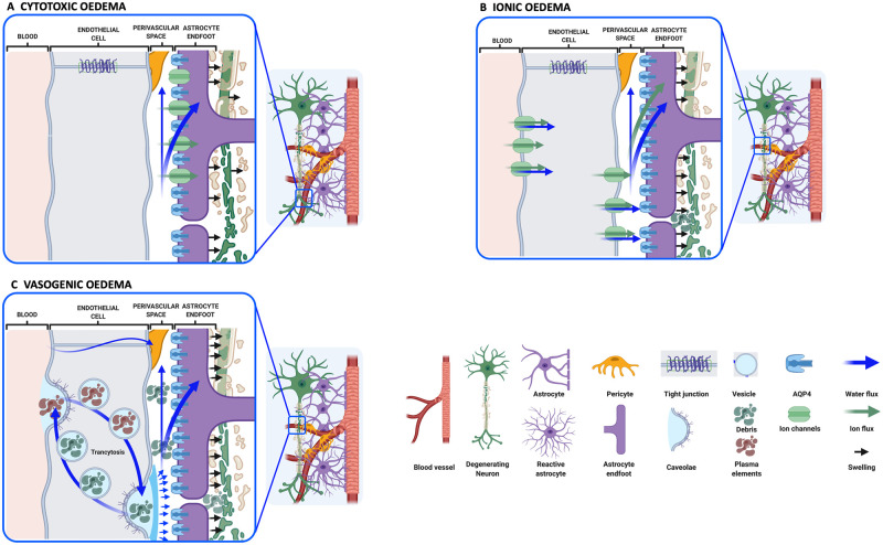
Aquaporin channels facilitate bidirectional water flow in all cells and tissues. AQP4 is highly expressed in astrocytes. In the CNS, it is enriched in astrocyte endfeet, at synapses, and at the glia limitans, where it mediates water exchange across the blood-spinal cord and blood-brain barriers (BSCB/BBB), and controls cell volume, extracellular space volume, and astrocyte migration. Perivascular enrichment of AQP4 at the BSCB/BBB suggests a role in glymphatic function. Recently, we have demonstrated that AQP4 localization is also dynamically regulated at the subcellular level, affecting membrane water permeability. Ageing, cerebrovascular disease, traumatic CNS injury, and sleep disruption are established and emerging risk factors in developing neurodegeneration, and in animal models of each, impairment of glymphatic function is associated with changes in perivascular AQP4 localization. CNS oedema is caused by passive water influx through AQP4 in response to osmotic imbalances. We have demonstrated that reducing dynamic relocalization of AQP4 to the BSCB/BBB reduces CNS oedema and accelerates functional recovery in rodent models. Given the difficulties in developing pore-blocking AQP4 inhibitors, targeting AQP4 subcellular localization opens up new treatment avenues for CNS oedema, neurovascular and neurodegenerative diseases, and provides a framework to address fundamental questions about water homeostasis in health and disease.

Keywords: neurodegeneration; regulation; traumatic brain and spinal cord injury; water channel.


Figures

Figure 1
Figure 1
AQP4 localization in the CNS. (A) AQP4 (blue) is located within astrocyte endfeet processes surrounding blood vessels in both brain tissue and the BBB. The inset shows the crystal structure of human AQP4 (PDB code 3GD8). AQP4 assembles as a tetramer with each monomer comprising six transmembrane helices and two half-helices (grey). The two half helices harbour the aquaporin signature motif (NPA) as well as part of the aromatic-arginine (ar/R) motif that functions as a selectivity filter. Within the pore, water molecules (red spheres) align in a single file. (B) AQP4 is localized at the astrocyte component of the tripartite synapse. During neurotransmission, neurons release mediators and neurotransmitters from synaptic nerve terminals (affecter cells) into the synaptic cleft to communicate with other neurons (effector cells). This synaptic activity induces an increase in intracellular Ca2+ concentration, which is accompanied by changed water and solute concentrations in astrocytes, leading to the release of glutamate and other gliotransmitters. This gliotransmission results in negative feedback to the presynaptic neurons to modulate neurotransmission. AQP4 plays an essential role in maintaining water homeostasis during this process. (C) In ventricles, AQPs are present within ependymal cells lining the brain-CSF interfaces (left inset). AQP4 is localized to the basolateral membrane of ependymal cells and the endfeet of contacting astrocytes (right inset). AQP1 (purple) is localized to the apical membrane of the choroid plexus epithelium., (D) CSF within the subarachnoid and cisternal spaces flows into the brain specifically via periarterial spaces and then exchanges with brain interstitial fluid facilitated by AQP4 water channels that are positioned within perivascular astrocyte endfoot processes.
Figure 2
Figure 2
The glymphatic pathway. The glymphatic system is a perivascular network that facilitates fluid exchange between the CSF and interstitial compartments, supporting the clearance of interstitial solutes. The function of the glymphatic system relies on perivascular astrocyte AQP4 expression. In the healthy young brain, AQP4 localizes to the astrocyte endfeet along the perivascular space (top left, arrows). In the context of ageing and Alzheimer’s disease, perivascular AQP4 levels are reduced while cellular AQP4 levels are increased (bottom left, arrows). The loss of AQP4 from perivascular astrocytic endfeet slows glymphatic clearance, which may accelerate amyloid-β accumulation and cognitive decline. The column on the right details specific findings from studies in rodents (top) and humans (bottom).
Figure 3
Figure 3
The pathogenesis of traumatic injury in the CNS. In the primary injury phase, the brain or spinal cord is injured following external insult. This primary injury results in mechanical damage to neurons, astrocytes, oligodendrocytes and blood vessels. A series of secondary injury cascades then occurs that potentiates the primary injury. In the earlier post-injury stages, damaged blood vessels may haemorrhage, resulting in ischaemia and release of inflammatory cytokines (e.g. TGF-β, TNF-α, IL-1, and IL-6). These cytokines attract blood-borne inflammatory cells such as neutrophils, macrophages and leucocytes, which act both to clear up cellular debris, but also cause further damage to healthy cells by enhancing local inflammation, eventually leading to neuronal loss from inflammatory damage and through Wallerian degeneration following oligodendrocyte death and demyelination. Damaged neurons may secrete free radicals, nitric oxide (NO), glutamate, and Ca2+, which further potentiate cellular damage by causing mitochondrial dysfunction leading to the loss of ATP, and by causing localized excitotoxicity. Collectively, these two events result in the loss of Na+/K+-ATPase activity and the loss of oxygen tension in astrocytes, which results in cytotoxic oedema through increased water absorption through AQP4 (blue). This is followed by ionic dysregulation, eventually leading to swelling via vasogenic oedema and cavity formation limited by the formation of a glial scar, which obstructs neuronal regrowth and enhances cell damage. Created using www.biorender.com.
Figure 4
Figure 4
Classification of CNS oedema. (A) Cytotoxic oedema is defined by astrocyte swelling (black arrows) followed by neuronal dendrite swelling. The net entry of water (blue arrows), most likely from the perivascular space, is caused by disruption of cellular ion homeostasis (green arrows) following hypoxic insult. (B) Ionic oedema is characterized by transcapillary sodium ion and anion fluxes associated with cellular uptake of ions from the perivascular CSF, and entry of water into the brain parenchyma. Astrocytes continue to be swollen (black arrows) by water from the perivascular space and the vascular compartment. Neuronal death produces cellular debris in the extracellular space (ECS). (C) Vasogenic oedema is a result of BBB dysfunction, possibly following ionic oedema. Increased transcytosis may contribute to the entry of plasma elements (brown), followed by water. Clearance of debris from the ECS produced by neuronal cell death may also occur by transcytosis (green). In some severe cases, the tight junctions between the endothelial cells are weakened leading to increased permeability of cerebral blood vessels to plasma components. Created using www.biorender.com.

References

    1. Rangel-Castilla L, Rangel-Castillo L, Gopinath S, Robertson CS.. Management of intracranial hypertension. Neurol Clin. 2008;26(2):521–541. - PMC - PubMed
    1. Nagelhus EA, Ottersen OP.. Physiological roles of aquaporin-4 in brain. Physiol Rev. 2013;93(4):1543–1562. - PMC - PubMed
    1. Mader S, Brimberg L.. Aquaporin-4 water channel in the brain and its implication for health and disease. Cells. 2019;8(2):90. - PMC - PubMed
    1. Manley GT, Binder DK, Papadopoulos MC, Verkman AS.. New insights into water transport and edema in the central nervous system from phenotype analysis of aquaporin-4 null mice. Neuroscience. 2004;129(4):983–991. - PubMed
    1. Verkman AS, Binder DK, Bloch O, Auguste K, Papadopoulos MC.. Three distinct roles of aquaporin-4 in brain function revealed by knockout mice. Biochim Biophys Acta. 2006;1758(8):1085–1093. - PubMed

Publication types