. 2021 Sep 9;13(17):20935-20961.
doi: 10.18632/aging.203485. Epub 2021 Sep 9.

Hyperbaric oxygen therapy alleviates vascular dysfunction and amyloid burden in an Alzheimer's disease mouse model and in elderly patients

Affiliations

Hyperbaric oxygen therapy alleviates vascular dysfunction and amyloid burden in an Alzheimer's disease mouse model and in elderly patients

Ronit Shapira et al. Aging (Albany NY). .

Abstract

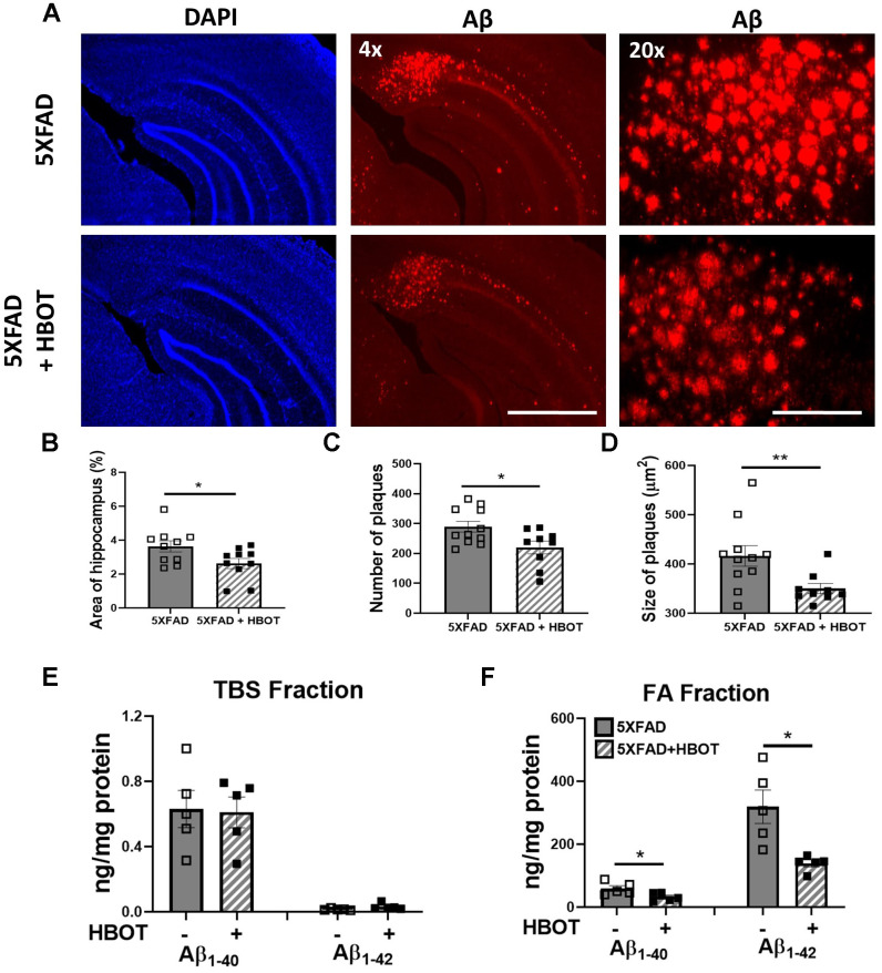
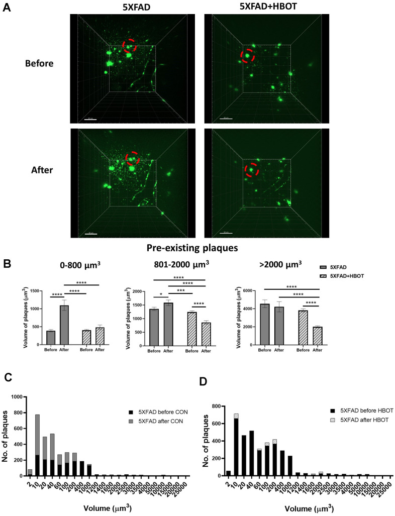
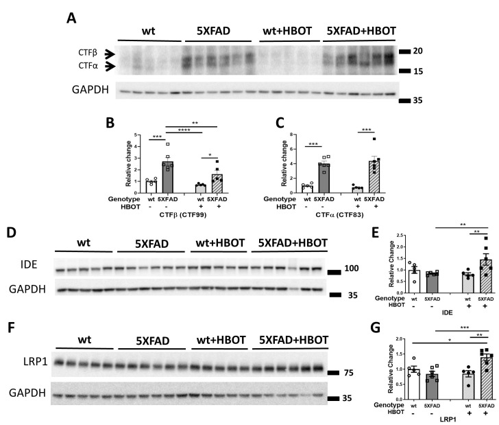
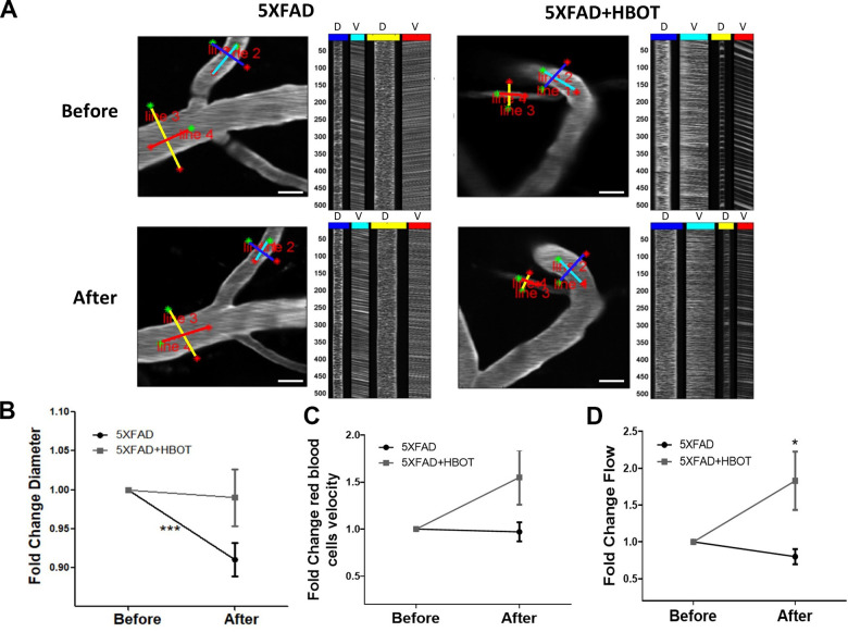
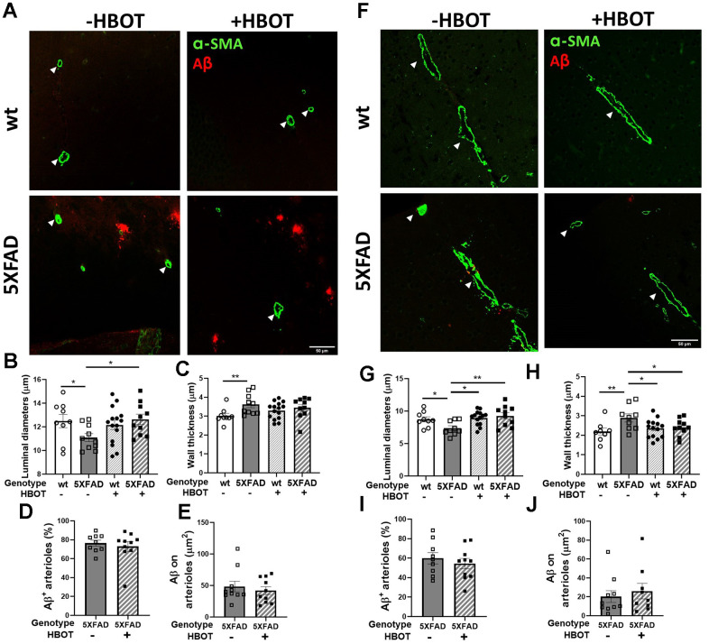
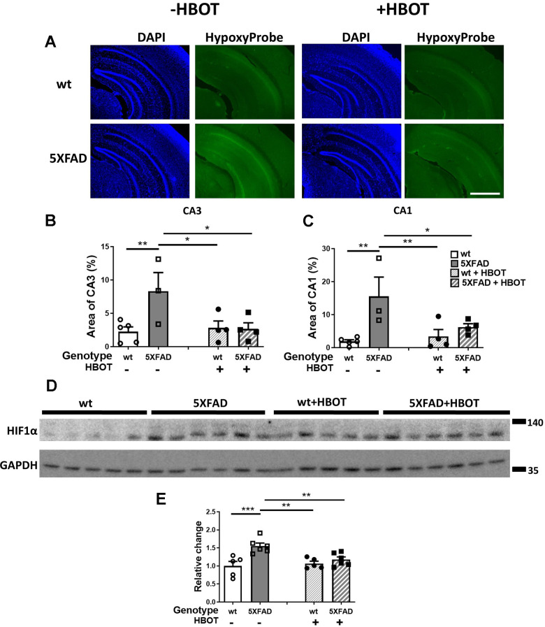
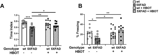
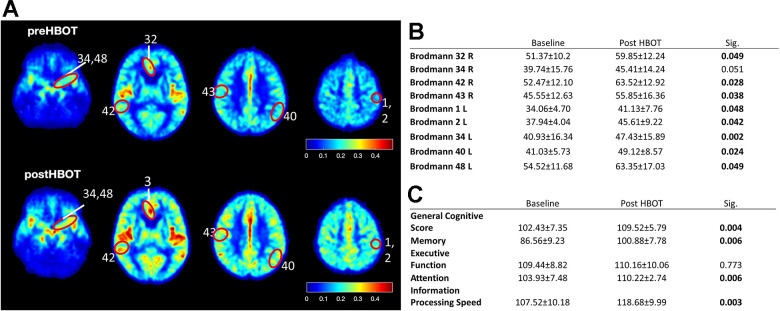
Vascular dysfunction is entwined with aging and in the pathogenesis of Alzheimer's disease (AD) and contributes to reduced cerebral blood flow (CBF) and consequently, hypoxia. Hyperbaric oxygen therapy (HBOT) is in clinical use for a wide range of medical conditions. In the current study, we exposed 5XFAD mice, a well-studied AD model that presents impaired cognitive abilities, to HBOT and then investigated the therapeutical effects using two-photon live animal imaging, behavioral tasks, and biochemical and histological analysis. HBOT increased arteriolar luminal diameter and elevated CBF, thus contributing to reduced hypoxia. Furthermore, HBOT reduced amyloid burden by reducing the volume of pre-existing plaques and attenuating the formation of new ones. This was associated with changes in amyloid precursor protein processing, elevated degradation and clearance of Aß protein and improved behavior of 5XFAD mice. Hence, our findings are consistent with the effects of HBOT being mediated partially through a persistent structural change in blood vessels that reduces brain hypoxia. Motivated by these findings, we exposed elderly patients with significant memory loss at baseline to HBOT and observed an increase in CBF and improvement in cognitive performances. This study demonstrates HBOT efficacy in hypoxia-related neurological conditions, particularly in AD and aging.

Keywords: Alzheimer's disease; amyloid burden; cerebral blood flow; hyperbaric oxygen therapy; vascular dysfunction.


Conflict of interest statement

CONFLICTS OF INTEREST: The authors declare that they have no conflicts of interest. AH, ES work for AVIV Scientific LTD. SE is a co-founder and shareholder at AVIV Scientific LTD.

Figures

Figure 1
Figure 1
HBOT reduces amyloid plaques in the hippocampal area of 6-month old 5XFAD mice. Amyloid plaques were visualized by immunostaining with anti-Aβ antibodies (4G8). (A) Representative images of Aβ in the hippocampal field of HBO-treated 5XFAD (n=10, lower panel) and control 5XFAD mice (n=10, upper panel); left and middle panels, x4 magnification, scale bar: 1000 μm; right panel, x20 magnification, scale bar: 200 μm. (B) Quantification of the percentage of hippocampal area occupied by plaques. (C) Number of plaques. (D) Mean size of plaques. (E, F) Soluble Aβ was initially extracted from hippocampi with TBS by ultracentrifugation and then insoluble Aβ was extracted with 70% formic acid (FA) after ultra-centrifugation. ELISA analysis of soluble (E) and insoluble (F) Aβ40 and Aβ42 in hippocampal lysates of HBO-treated 5XFAD and control 5XFAD mice (n = 5/group). (B, C, F) -t-test, (D, F)- welch correction t-test. Values represent means ± SEM. *P < 0.05, ** P < 0.01.
Figure 2
Figure 2
HBOT reduces the population of newly-formed plaques and reduces the volume of pre-existing plaques. Amyloid plaques were visualized in vivo using two-photon microscopy imaging in live animals by injecting methoxy-X04 24 h before every imaging session. (A) Representative images of plaques in the somatosensory cortex of HBO-treated 5XFAD (n=4, right panel) and control 5XFAD mice (n=3, left panel) before (upper panel) and after 1 month of treatment (lower panel); red circles indicate the change in specific plaques, scale bar: 50 μm. (B) Analysis of the volume of pre-existing plaques before and after each treatment in the same animal, categorized according to initial plaque size. (C, D) Distribution of plaque populations by volume in control 5XFAD (C; before: N=1619, after: N=3180) and HBO-treated 5XFAD mice (D; before: N=3425, after: N=3524). Two-way ANOVA with repeated measures and post-hoc Fisher LSD tests were performed. Values represent means ± SEM. *P < 0.05, *** P < 0.001, **** P < 0.0001.
Figure 3
Figure 3
HBOT reduces abnormal processing of APP and attenuates Aβ degradation and clearance in 5XFAD mice. (A) Representative immunoblot assays of the carboxyl-terminal fragment (CTF)β and CTFα. (B, C) Quantification of western blots in (A), presented as percentages of wt control, normalized to GAPDH levels (n = 5–6/group). (DG) Representative immunoblot assays of IDE protein (D) and LRP1 in (F). (E, G) Quantification of western blots in (D, F), respectively, presented as percentages of wt controls, normalized to GAPDH levels (n = 5-6/group). Two-way ANOVA and post-hoc Fisher LSD tests were performed. Values represent means ± SEM. * P < 0.05, ** P<0.01, *** P < 0.001, **** P < 0.0001.
Figure 4
Figure 4
HBOT alleviates the reduction in vessel diameter in 5XFAD mice and increases blood flux.In vivo two-photon microscopy imaging and measurements of diameter and velocity in blood vessels of the somatosensory cortex in 5XFAD mice using spatially optimized line scans. (A) Representative images of fluorescently stained vessels of the somatosensory cortex of an HBO-treated 5XFAD mouse (right panel) and a control 5XFAD mouse (left panel) before (upper panel) and after a month of treatment (lower panel). Line scan patterns are superimposed on the vessels. Lines of the scan path along the length were used to calculate RBC velocity (V), while lines across the diameter of the vessels were used to calculate diameter (D). The line scans generated from the path can be stacked sequentially as a function of time to produce a raw cascade image (right of each image). Vessel diameter was calculated as the full width at half-maximum of a time average of several scans across the width of a vessel. RBC velocity was calculated from the angle of the RBC streaks. (BD) Quantification of vessel diameter (B), RBC velocity in the blood vessels (C) and RBC flow (D), normalized to each treatment group baseline value. Paired t-tests and student t-tests were performed. Values represent means ± SEM.* P < 0.05, *** P < 0.001.
Figure 5
Figure 5
HBOT attenuates arteriolar luminal diameter but not amyloid deposition around arterioles in 6-month old 5XFAD mice. Arterioles were visualized using immunostaining with anti-SMA antibody while vascular amyloid deposition was visualized using anti-Aβ antibody (4G8). (A, F) Representative images of arterioles and Aβ in hippocampal (A) and cortical fields (F) of HBO-treated wt (n=9, upper right panels) and 5XFAD mice (n=10, lower right panels) and control wt (n=9, upper left panels) and 5XFAD mice (n=10, lower left panels) (x40 magnification, scale bar: 50 μm). White arrows show hippocampal and cortical arterioles. (BE) and (GJ), Quantification of arteriolar luminal diameters (B, G), arteriolar wall thickness (C, H) and percentages of arterioles that stained positive for Aβ (D, I) and Aβ deposition area around arterioles (E, J) in the hippocampal (BE) and cortical fields (GJ). Two-way ANOVA and post-hoc Fisher LSD tests were performed. Values represent means ± SEM. * P < 0.05, ** P < 0.01.
Figure 6
Figure 6
HBOT reduces hypoxia and HIF1α transcription factor levels in the hippocampal area of 6-month old 5XFAD mice. (A) Uptake of Hypoxyprobe by low oxygen-bearing cells was visualized by immunostaining. Representative images of the presence of hypoxia in the hippocampal field of HBO-treated wt (n=4, right upper panel) and 5XFAD mice (n=4, right lower panel), and control wt (n=5, left upper panel) and 5XFAD mice (n=3, left lower panel); x4 magnification, scale bar: 1000 μm. (B, C) Quantification of the percentage of the CA3 (B) and CA1 (C) areas presenting Hypoxyprobe-related fluorescence. (D) Western blots of HIF-1α from hippocampi extracted from HBO-treated and control 5XFAD mice and wt littermates. (E) Quantification of Western blots in (D), presented as percentage of wt control, normalized to GAPDH levels (n = 4–5/group). Two-way ANOVA and post-hoc Fisher LSD tests were performed. Values represent means ± SEM. *P < 0.05, ** P < 0.01, *** P < 0.001.
Figure 7
Figure 7
HBOT improves performance of 5XFAD mice in cognitive tasks. (A) In the Y-maze test, HBO-treated 5XFAD mice showed better spatial memory as reflected in the time index, which is displayed as the ratio (novel /novel + familiar) to time in each arm. (B) In the trace fear conditioning assay, mice underwent conditioning involving 6 rounds of tone-shock pairing with a trace interval. On the following day, the mice were exposed to the same context with no exposure to tone or shock. Results of contextual freezing are expressed as the percent of total time spent frozen in the training context. Two-way ANOVA with/without repeated measures and post-hoc Fisher LSD tests were performed. Values represent means ± SEM. *P < 0.05, **P < 0.01, *** P < 0.001.
Figure 8
Figure 8
CBF and cognitive function are improved following HBOT of patients. CBF and cognitive functions of six patients suffering from memory decline at baseline and following 60 HBOT sessions. (A) Average normalized CBF maps (DSC) at baseline and post-HBOT. (B) Significant average CBF changes in Brodmann areas at baseline and post-HBOT. (C) Average cognitive domain scores (Neurotrax) at baseline and post-HBOT.

References

    1. Roher AE, Debbins JP, Malek-Ahmadi M, Chen K, Pipe JG, Maze S, Belden C, Maarouf CL, Thiyyagura P, Mo H, Hunter JM, Kokjohn TA, Walker DG, et al.. Cerebral blood flow in Alzheimer’s disease. Vasc Health Risk Manag. 2012; 8:599–611. 10.2147/VHRM.S34874 - DOI - PMC - PubMed
    1. Wolters FJ, Zonneveld HI, Hofman A, van der Lugt A, Koudstaal PJ, Vernooij MW, Ikram MA, and Heart-Brain Connection Collaborative Research Group. Cerebral Perfusion and the Risk of Dementia: A Population-Based Study. Circulation. 2017; 136:719–28. 10.1161/CIRCULATIONAHA.117.027448 - DOI - PubMed
    1. Akoudad S, Wolters FJ, Viswanathan A, de Bruijn RF, van der Lugt A, Hofman A, Koudstaal PJ, Ikram MA, Vernooij MW. Association of Cerebral Microbleeds With Cognitive Decline and Dementia. JAMA Neurol. 2016; 73:934–43. 10.1001/jamaneurol.2016.1017 - DOI - PMC - PubMed
    1. Santos CY, Snyder PJ, Wu WC, Zhang M, Echeverria A, Alber J. Pathophysiologic relationship between Alzheimer’s disease, cerebrovascular disease, and cardiovascular risk: A review and synthesis. Alzheimers Dement (Amst). 2017; 7:69–87. 10.1016/j.dadm.2017.01.005 - DOI - PMC - PubMed
    1. Vinters HV. Cerebral amyloid angiopathy. A critical review. Stroke. 1987; 18:311–24. 10.1161/01.str.18.2.311 - DOI - PubMed

Publication types

Substances