. 2022 Jan 6;30(1):114-128.e9.
doi: 10.1016/j.str.2021.08.007. Epub 2021 Sep 8.

Molecular mechanisms underlying the role of the centriolar CEP164-TTBK2 complex in ciliopathies

Affiliations

Molecular mechanisms underlying the role of the centriolar CEP164-TTBK2 complex in ciliopathies

Ivan Rosa E Silva et al. Structure. .

Abstract

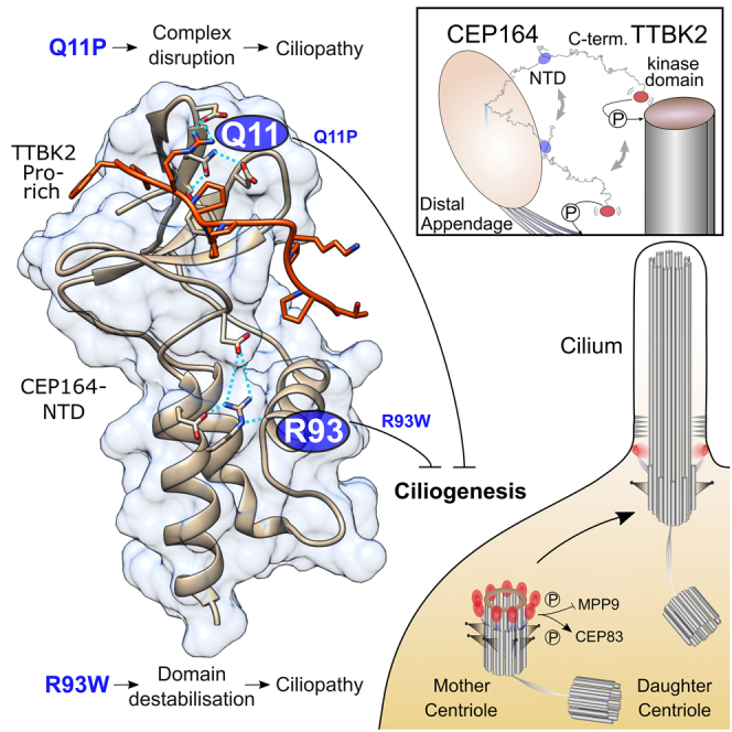
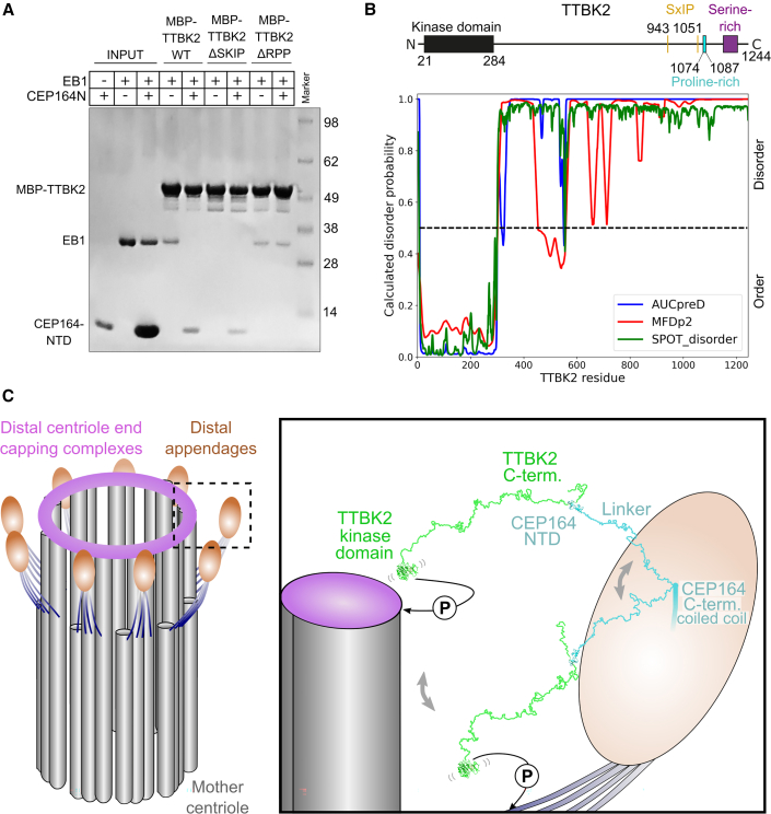
Cilia formation is essential for human life. One of the earliest events in the ciliogenesis program is the recruitment of tau-tubulin kinase 2 (TTBK2) by the centriole distal appendage component CEP164. Due to the lack of high-resolution structural information on this complex, it is unclear how it is affected in human ciliopathies such as nephronophthisis. Furthermore, it is poorly understood if binding to CEP164 influences TTBK2 activities. Here, we present a detailed biochemical, structural, and functional analysis of the CEP164-TTBK2 complex and demonstrate how it is compromised by two ciliopathic mutations in CEP164. Moreover, we also provide insights into how binding to CEP164 is coordinated with TTBK2 activities. Together, our data deepen our understanding of a crucial step in cilia formation and will inform future studies aimed at restoring CEP164 functionality in a debilitating human ciliopathy.

Keywords: CEP164; TTBK2; basal body; centriole; centrosome; cilia; ciliogenesis; ciliopathy; distal appendage; nephronophthisis.


Conflict of interest statement

Declaration of interests The authors declare no competing interests.

Figures

None
Graphical abstract
Figure 1
Figure 1
High-resolution structure of CEP1641−109 that interacts with TTBK21074−1087 with low micromolar affinity (A) Domain organization of human CEP164 and TTBK2. cc, coiled coil. Amino acid residue numbers are indicated. (B) The WW domain of CEP164 and the proline-rich region 1,074–1,087 in TTBK2 are necessary for the CEP164-TTBK2 interaction. Western blots showing pull-down experiments with lysates from cells expressing 3xFLAG-tagged CEP164 and 3xHA-tagged TTBK2 constructs. Left: pull-down with 3xFLAG-CEP164 and 3xHA-TTBK2 or 3xHA-TTBK2 carrying a deletion of its proline-rich region 1,074–1,087 (ΔPro.). Right: pull-down with 3xFLAG-CEP164 or 3xFLAG-CEP164 carrying a deletion containing its WW domain (ΔWW) and 3xHA-TTBK2. TCL, total cell lysate. (C) Recombinant CEP1641−109 and TTBK21074−1087 interact with low micromolar affinity. Typical ITC of a synthetic TTBK21074−1087 peptide and recombinant human CEP1641−109 constructs carrying the indicated mutations, at 25°C. The resulting dissociation constants (KD) are indicated as an average (±SD) from three to five independent measurements. (D) Top left: ribbon representation of the structure of CEP1641−109, rainbow-colored from N- to C-terminus, in complex with a camelid nanobody (in gray). Rotation, as indicated. Consecutive alpha helices (α), beta sheets (β), and linkers (L) are labeled. Dotted boxes labeled from 1 to 3 indicate the regions shown magnified in the three panels below (1 and 2) or to the right (3). Bottom: detailed view of the CEP1641−109 regions containing residues R93 (box 1) or Q11 (box 2). These residues are mutated to W or P, respectively, in ciliopathies. Yellow dotted lines indicate hydrogen bonds. Selected residues are shown as sticks and are labeled. bb, backbone. Top right, ribbon representation of the region indicated by box 3, colored according to ConSurf (Ashkenazy et al., 2016) conservation scores from variable (cyan) to conserved (burgundy). Key residues in the interface region between the WW domain and the helical bundle are shown as sticks and are labeled.
Figure 2
Figure 2
The isolated helical bundle but not the WW domain of CEP1641−109 folds stably in solution (A) Left, NMR structure ensemble of CEP1641−109 superposed to its WW domain (CEP16462−83), rainbow-colored from N (blue) to C terminus (red). Right, corresponding view of the superposed crystal structure of nanobody-bound CEP1641−109 (nanobody in gray). The corresponding RMSD values (±SD) were 0.7 ± 0.2 Å and 0.9 ± 0.1 Å, respectively. Consecutive alpha-helices (α), beta-sheets (β), and linkers (L) are labeled. The vectors used to calculate the helical bundle-WW domain orientation angles (in C) are indicated. The dotted box designates the region shown magnified below. Bottom, detailed view of the CEP1641−109 region contacted by the nanobody CDR3 loop. (B) Similar to (A), but CEP1641-109 NMR structures superposed to their helical bundle (CEP16426−55, 87-103). The corresponding RMSD values (±SD) were 0.5 ± 0.1 Å and 0.8 ± 0.1 Å, respectively. (C) Measured angles between the helical bundle and the WW domain in the NMR and crystal structures as indicated in (A). The average value across the NMR ensemble was 107° ± 7°. The individual angle values in each ensemble member are overlaid to a box-and-whisker plot. The whiskers represent the maximum and the minimum measured values, while the box ranges between the first and the third quartiles; the median value is also indicated. (D) Top: ribbon representation of the CEP1641−109 structure (in green). The ΔHelices and ΔWW panels indicate the construct deletions of the helical bundle or WW domain, respectively, by omitting the corresponding structural regions. Dotted black lines highlight the parts that were directly spliced together in the constructs. Alpha helices (α), beta sheets (β), and linkers (L) are labeled. Bottom: CD analyses of recombinant CEP1641−109 WT as well as the corresponding ΔHelices and ΔWW constructs. Left: buffer-subtracted CD spectra at 25°C. Right: CD-based thermal melting analysis recorded at 222 nm. (E) The helical bundle of CEP164 is necessary for the CEP164-TTBK2 interaction. Western blot showing a pull-down experiment with lysates from cells expressing 3xHA-TTBK2 and 3xFLAG tagged CEP164 or 3xFLAG-CEP164 carrying a deletion of its helical bundle. ΔHel., construct as indicated by ΔHelices in (D). TCL, total cell lysate.
Figure 3
Figure 3
High-resolution structure of the CEP1641−109-TTBK21074−1087 complex that is compromised by the ciliopathy-associated mutations Q11P and R93W in CEP164 (A) Left: ribbon representation of the structure of CEP1641−109 (green) in complex with TTBK21074−1087 (red). The structure of the bound camelid nanobody used for co-crystallization is omitted for optical clarity. Consecutive alpha-helices (α), beta-sheets (β), and linkers (L) are labeled. The dotted box is shown magnified to the right. Right: detailed view of the interface region between CEP1641−109 and TTBK21074−1087. Key interface residues are shown as sticks and are labeled. Yellow dotted lines indicate hydrogen bonds. (B) Several interfacial residues of the CEP164 WW domain are critical for its interaction with TTBK2. Western blot showing a pull-down experiment with recombinant GST or GST-CEP1641−109, WT or the indicated mutants, and lysates from cells expressing C-terminally GFP-tagged TTBK21074−1087. Below the blot, the band intensities of the eluates (as percentage of the WT level) are shown. (C) Several residues within the CEP164-binding region of TTBK2 are essential for its interaction with CEP164 and define a CEP164-binding motif. Western blot showing a pull-down experiment with recombinant GST or GST-CEP1641−109 and lysates from cells expressing C-terminally GFP-tagged TTBK21074−1087 constructs. TTBK2 constructs carried single alanine mutations as indicated above the blot. Below the blot, the band intensities of the eluates (as percentage of the WT level) are shown (±SD, n = 3). (D and E) The R93W, but not the Q11P mutation decrease the thermostability of the NTD of CEP164. (D) Buffer-subtracted CD spectra of recombinant CEP1641−109 WT as well as its Q11P and R93W mutants at 7°C and 25°C. (E) CD-based thermal melting analysis of CEP1641−109 WT, Q11P, and R93W at 222 nm. (F) Both Q11P and R93W nephronophthisis mutations in CEP1641−109 compromise the interaction with TTBK21074−1087 at 4°C. Western blot showing a pull-down experiment with recombinant GST or GST-CEP1641−109, WT, Q11P, or R93W and lysates from cells expressing C-terminally GFP-tagged TTBK21074−1087. Below the blot, the band intensities of the eluates (as percentage of the WT level) are shown (±SD, n = 3). (G and H) The CEP164 Q11P and R93W ciliopathy mutants localize to mother centrioles but do not efficiently rescue cilia formation in CEP164-null cells. (G) Immunofluorescence staining of hTERT RPE-1 CEP164-null cells rescued with the indicated, transfected 3xFLAG-tagged CEP164 constructs (green). Primary cilia were detected by ARL13b staining (red). CETN1 was used to visualize centrosomes (blue). Scale bar: 5 μm. (H) Quantification of the percentage of ciliated cells (mean percentage of ciliated cells ±SEM). Statistical significance between the groups was analyzed by one-way ANOVA with a Holm-Sidac multiple comparison post hoc test; number of cells, NWT = 66, NQ11P = 58, NR93W = 55, and Nuntransfected = 57; p = 0.02, ∗∗∗p ≤ 0.0005.
Figure 4
Figure 4
CEP164-NTD binding to TTBK2 inhibits the TTBK2-EB1 interaction (A) CEP1641−109 and EB1 compete for binding to MBP-TTBK21033−1087. Coomassie-stained SDS-PAGE gel showing pull-down experiments with purified MBP-TTBK21033−1087 (WT, EB1-binding-deficient TTBK21051−1054 SKIP to AAAA or CEP164-NTD-binding-deficient TTBK21076−1078 RPP to AAA) with EB1 in the presence of an excess of CEP1641−109. (B) Per-residue disorder probabilities of TTBK2 as calculated by different disorder prediction algorithms. Values above 0.5 (dashed line) indicate disorder. The location of the kinase domain and selected sequence features of TTBK2 are indicated above the plot. (C) Model of the TTBK2-CEP164 architecture at the distal appendages. Left: scheme of the mother centriole with distal appendages based on electron microscopy tomography segmentation analysis (Bowler et al., 2019). Subdistal appendages are omitted for clarity. Right: close-up view of a single distal appendage region. CEP164 is indicated in cyan, the bound TTBK2 is shown in green. The connecting linkers between CEP164-NTD and the CEP164 coiled-coil domain and between the CEP164-bound TTBK21074−1084 region and the N-terminal TTBK2 kinase domain (PDB: 6vrf) (Bao et al., 2021) are drawn in an extended and open conformation. Based on disorder predictions and the paucity of predicted secondary structure elements, we propose that these linkers are largely flexible (Figures 4B and S6B). This flexibility would allow the TTBK2-CEP164-NTD complex to sample larger areas of the distal appendage region and would enable TTBK2 to reach its phosphorylation substrates at (MPP9) or close to (CEP83) the distal centriole end. A circled “P” indicates a phosphorylation event by TTBK2.

Comment in

References

    1. Afonine P.V., Grosse-Kunstleve R.W., Echols N., Headd J.J., Moriarty N.W., Mustyakimov M., Terwilliger T.C., Urzhumtsev A., Zwart P.H., Adams P.D. Towards automated crystallographic structure refinement with phenix.refine. Acta Crystallogr. D Biol. Crystallogr. 2012;68:352–367. - PMC - PubMed
    1. Aragón E., Goerner N., Zaromytidou A.I., Xi Q., Escobedo Al., Massague J., Macias M.J. A Smad action turnover switch operated by WW domain readers of a phosphoserine code. Genes Dev. 2011;25:1275–1288. doi: 10.1101/gad.2060811. - DOI - PMC - PubMed
    1. Argentaro A., Yang J.C., Chapman L., Kowalczyk M.S., Gibbons R.J., Higgs D.R., Neuhaus D., Rhodes D. Structural consequences of disease-causing mutations in the ATRX-DNMT3-DNMT3L (ADD) domain of the chromatin-associated protein ATRX. Proc. Natl. Acad. Sci. U S A. 2007;104:11939–11944. - PMC - PubMed
    1. Ashkenazy H., Abadi S., Martz E., Chay O., Mayrose I., Pupko T., Ben-Tal N. ConSurf 2016: an improved methodology to estimate and visualize evolutionary conservation in macromolecules. Nucleic Acids Res. 2016;44:W344–W350. - PMC - PubMed
    1. Bao C., Bajrami B., Marcotte D.J., Chodaparambil J.V., Kerns H.M., Henderson J., Wei R., Gao B., Dillon G.M. Mechanisms of regulation and diverse activities of tau-tubulin kinase (TTBK) isoforms. Cell. Mol. Neurobiol. 2021;41:669–685. doi: 10.1007/s10571-020-00875-6. - DOI - PMC - PubMed

Publication types

LinkOut - more resources