. 2021 Sep 2;26(17):5352.
doi: 10.3390/molecules26175352.

An Evaluation of Understudied Phytocannabinoids and Their Effects in Two Neuronal Models

Affiliations

An Evaluation of Understudied Phytocannabinoids and Their Effects in Two Neuronal Models

Alex Straiker et al. Molecules. .

Abstract

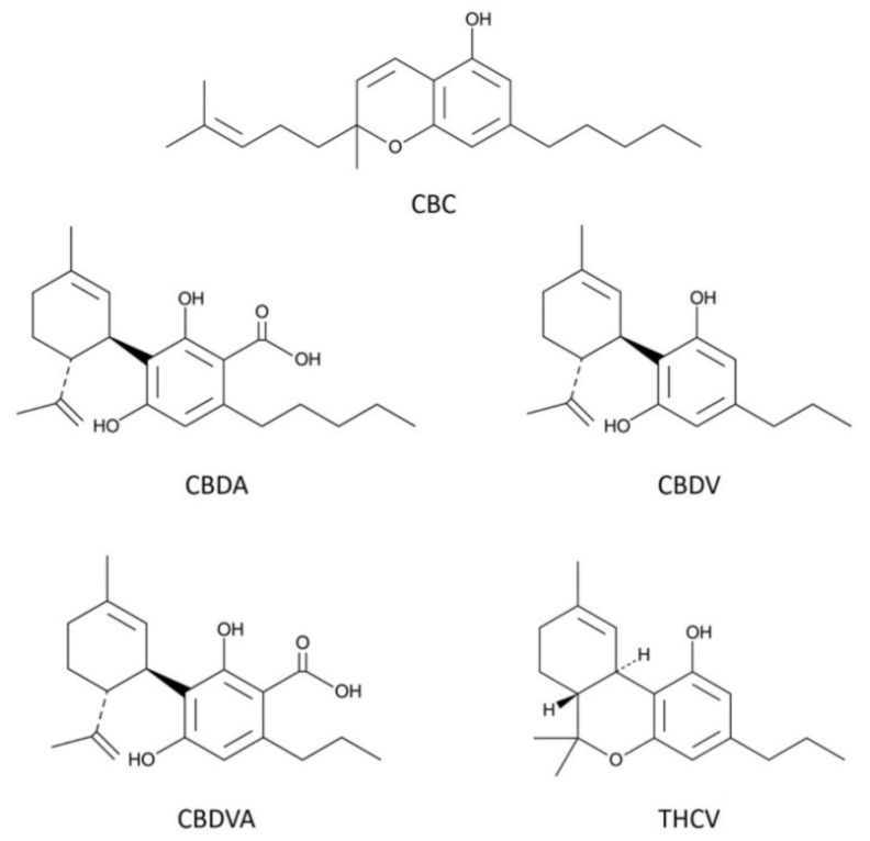
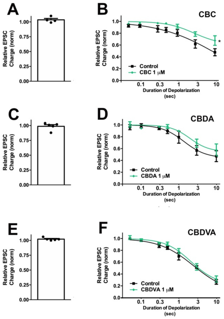
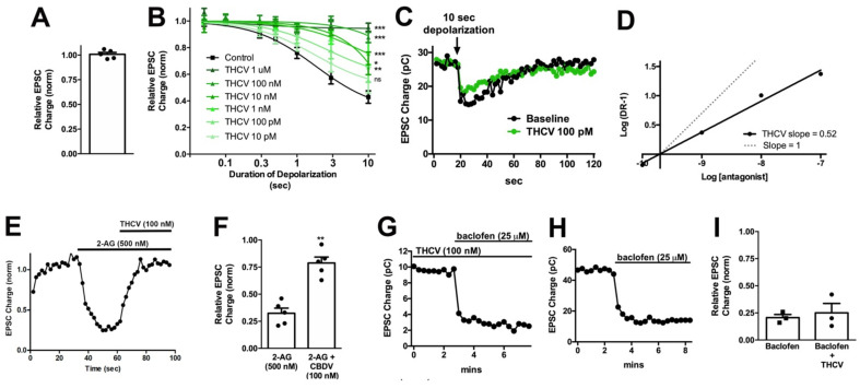
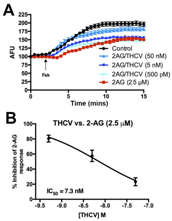
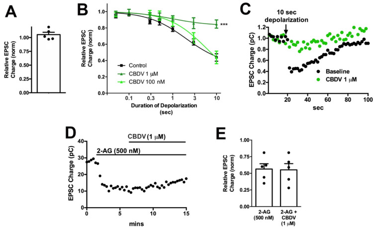
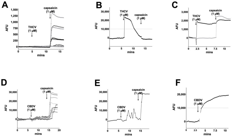
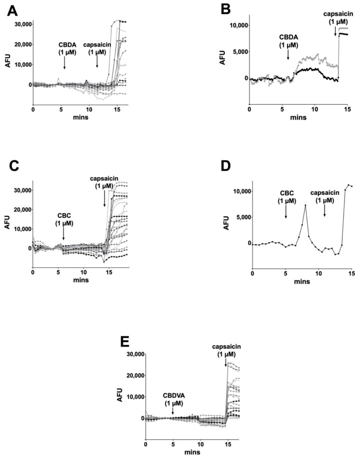
Cannabis contains more than 100 phytocannabinoids. Most of these remain poorly characterized, particularly in neurons. We tested a panel of five phytocannabinoids-cannabichromene (CBC), cannabidiolic acid (CBDA), cannabidivarin (CBDV), cannabidivarinic acid (CBDVA), and Δ9-tetrahydrocannabivarin (THCV) in two neuronal models, autaptic hippocampal neurons and dorsal root ganglion (DRG) neurons. Autaptic neurons expressed a form of CB1-dependent retrograde plasticity while DRGs expressed a variety of transient receptor potential (TRP) channels. CBC, CBDA, and CBDVA had little or no effect on neuronal cannabinoid signaling. CBDV and THCV differentially inhibited cannabinoid signaling. THCV inhibited CB1 receptors presynaptically while CBDV acted post-synaptically, perhaps by inhibiting 2-AG production. None of the compounds elicited a consistent DRG response. In summary, we find that two of five 'minor' phytocannabinoids tested antagonized CB1-based signaling in a neuronal model, but with very different mechanisms. Our findings highlight the diversity of potential actions of phytocannabinoids and the importance of fully evaluating these compounds in neuronal models.

Keywords: cannabichromene; cannabidiolic acid; cannabidivarin; cannabidivarinic acid; phytocannabinoids; tetrahydrocannabivarin.


Conflict of interest statement

The authors declare no conflict of interest.

Figures

Figure 1
Figure 1
Structures of phytocannabinoids examined in the current study. The figure shows chemical structures of the five phytocannabinoids used in this study; cannabichromene (CBC), cannabidiolic acid (CBDA), cannabidivarin (CBDV), cannabidivarinic acid (CBDVA), and Δ9-tetrahydrocannabivarin (THCV).
Figure 2
Figure 2
CBC modestly inhibits CB1 signaling in autaptic hippocampal neurons, while CBDA and CBDVA are without effect. (A) CBC, (C) CBDA, and (E) CBDVA have no direct effect on EPSCs. (B) CBC modestly inhibits maximal DSE. (D,F) CBDA and CBDV do not have a significant effect on DSE-mediated inhibition of EPSCs. *, p < 0.05, paired t-test for 10 s inhibition, drug vs. baseline.
Figure 3
Figure 3
THCV potently inhibits presynaptic CB1 responses in autaptic neurons. (A) THCV (1 μM) has no direct effect on EPSCs. (B) THCV concentration-dependently reduces DSE inhibition of EPSCs, with significant effects even at 100 pM. *, p < 0.05, ***, p < 0.005 one-way ANOVA with Dunnett’s post hoc vs. control. (C) Sample DSE responses before and after treatment with 1 μM THCV. (D) A Schild analysis shows that the Schild slope is less than 1. (E) Sample time course showing reversal of 2-AG inhibition by THCV (100 nM). (F) Summarized data showing that THCV reverses 2-AG action. **, p < 0.01 by paired t-test. (G,H) Sample time courses showing that baclofen (25 μM) responses are similar with and without pre-treatment with THCV (100 nM). (I) Summarized data for baclofen/THCV vs. baclofen alone. p > 0.05 unpaired t-test.
Figure 4
Figure 4
THCV inhibits 2-AG-mediated inhibition of adenylyl cyclase, but less potently than inhibition of neurotransmission. (A) Sample time courses from one set of experiments showing effects of drug combinations on forskolin-induced increases in cAMP in HEK293 cells transfected with mCB1 and the pink Flamindo cAMP indicator. (B) Summary cAMP responses show a concentration-dependent inhibition of 2-AG, with an IC50 of 7.3 nM.
Figure 5
Figure 5
CBDV inhibits DSE post-synaptically in autaptic neurons.; (A) CBDV (1 μM) has no direct effect on EPSC; (B) CBDV blocks DSE at 1 μM but not at 100 nM; (C) sample DSE responses before and after treatment with 1 μM CBDV; (D) sample time course showing non-reversal of 2-AG inhibition by CBDV (1 μM). (E) Summarized data showing that CBDV (1 μM) does not reverse the effect of 2-AG (500 nM). ***, p < 0.001, one-way ANOVA with Dunnett’s post-hoc test.
Figure 6
Figure 6
THCV and CBDV calcium responses in DRGs. (A) THCV (1 μM) rarely induced a calcium response in DRGs, while the TRPV1 agonist capsaicin (1 μM) induced responses in a large subset of DRGs. (B) In a small number of cells (~3%), THCV induced a desensitizing current. (C) A few cells (~3%) showed sustained responses to THCV. (D) CBDV (1 μM) did not typically induce a calcium response in DRGs. (E) In a few cells (~3%), CBDV appeared to increase spontaneous Ca transients. (F) A few cells (~4%) had a sustained calcium response to CBDV. AFU, arbitrary fluorescence units.
Figure 7
Figure 7
CBDA, CBC, and CBDVA rarely increased intracellular calcium in DRGs. (A) Sample time course shows calcium response in DRGs after treatment with CBDA (1 μM) followed by TRPV1 agonist capsaicin (1 μM), (B) on rare occasions (2%) DRG neurons responded to CBDA, (C) Sample time course shows calcium response in DRGs after treatment with CBC (1 μM) followed by capsaicin (1 μM), (D) on rare occasions (~1%) brief responses were seen after CBC treatment. (E) CBDVA failed to induce calcium responses in any cells tested.

References

    1. Elsohly M.A., Slade D. Chemical constituents of marijuana: The complex mixture of natural cannabinoids. Life Sci. 2005;78:539–548. doi: 10.1016/j.lfs.2005.09.011. - DOI - PubMed
    1. Gaoni Y., Mechoulam R. Isolation, structure and partial synthesis of an active constituent of hashish. J. Am. Chem. Soc. 1964;86:1646–1647. doi: 10.1021/ja01062a046. - DOI
    1. Howlett A.C., Barth F., Bonner T.I., Cabral G., Casellas P., Devane W.A., Felder C.C., Herkenham M., Mackie K., Martin B.R., et al. International Union of Pharmacology. XXVII. Classification of cannabinoid receptors. Pharmacol. Rev. 2002;54:161–202. doi: 10.1124/pr.54.2.161. - DOI - PubMed
    1. Matsuda L.A., Lolait S.J., Brownstein M.J., Young A.C., Bonner T.I. Structure of a cannabinoid receptor and functional expression of the cloned cDNA. Nature. 1990;346:561–564. doi: 10.1038/346561a0. - DOI - PubMed
    1. Munro S., Thomas K.L., Abu-Shaar M. Molecular characterization of a peripheral receptor for cannabinoids. Nature. 1993;365:61–65. doi: 10.1038/365061a0. - DOI - PubMed

LinkOut - more resources