. 2021 Aug 31;14(17):4987.
doi: 10.3390/ma14174987.

Characterization and Cytotoxicity Comparison of Silver- and Silica-Based Nanostructures

Affiliations

Characterization and Cytotoxicity Comparison of Silver- and Silica-Based Nanostructures

Elżbieta Adamska et al. Materials (Basel). .

Abstract

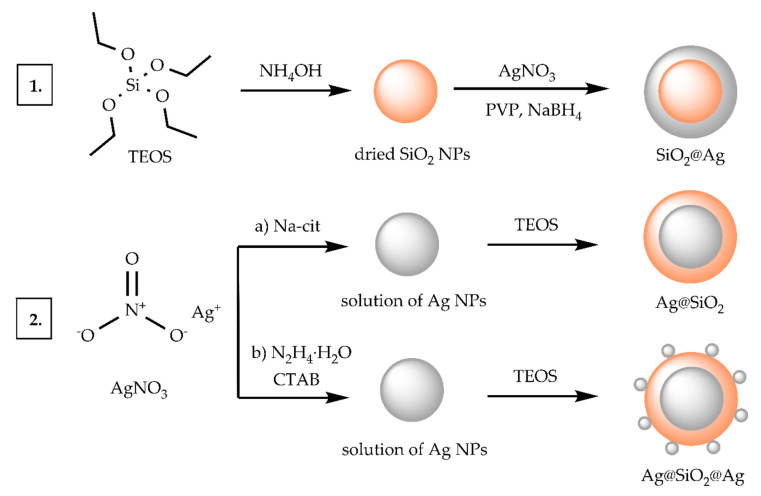
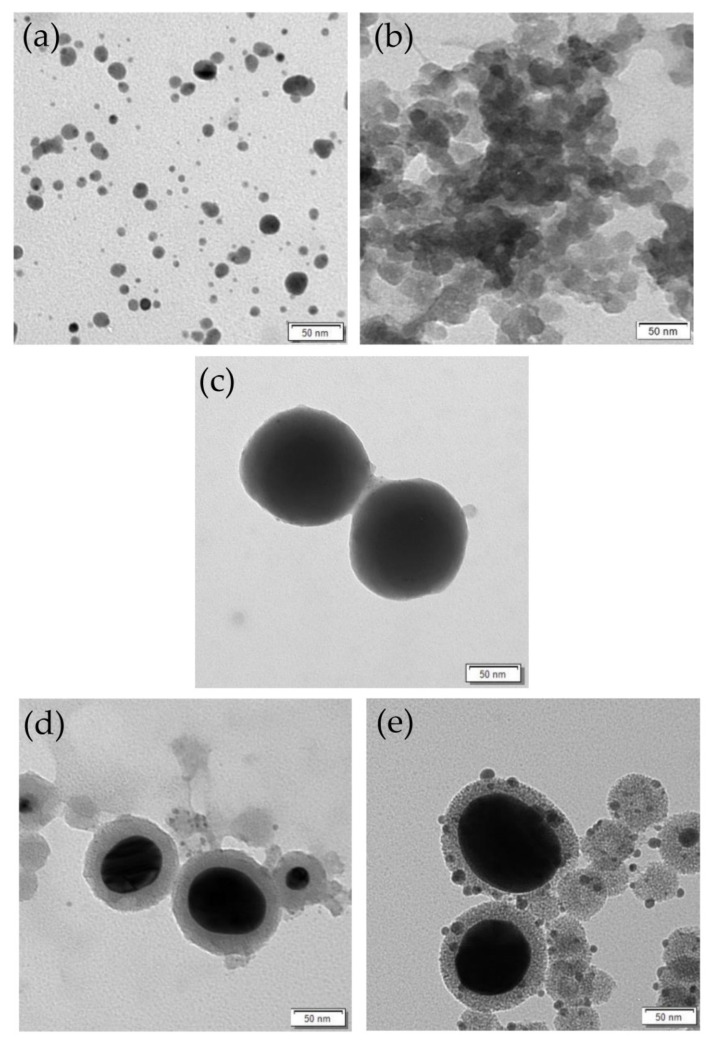
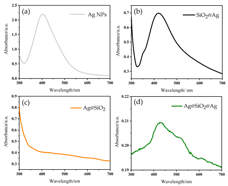
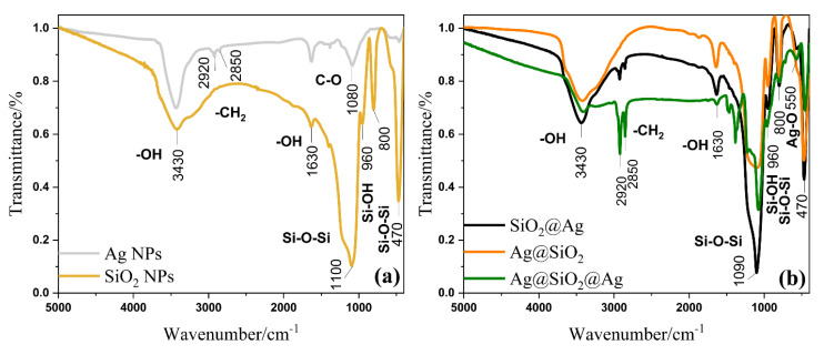
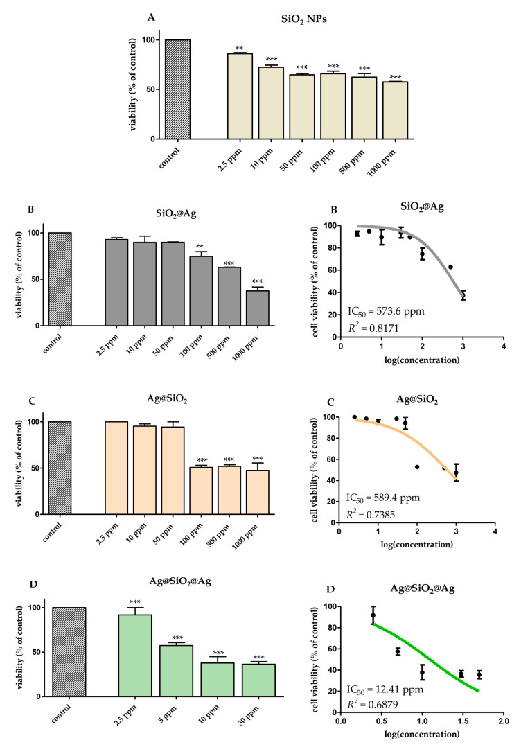
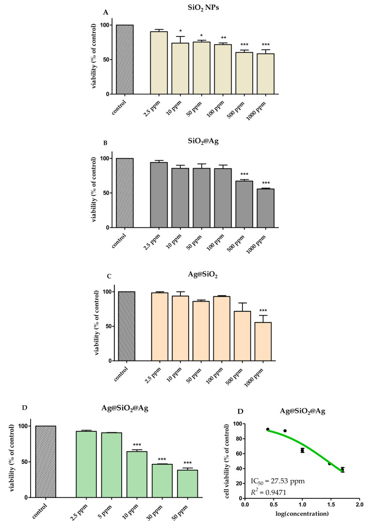
Core-shell structures are the most common type of composite material nanostructures due to their multifunctional properties. Silver nanoparticles show broad antimicrobial activity, but the safety of their utilization still remains an issue to tackle. In many applications, the silver core is coated with inorganic shell to reduce the metal toxicity. This article presents the synthesis of various materials based on silver and silica nanoparticles, including SiO2@Ag, Ag@SiO2, and sandwich nanostructures-Ag@SiO2@Ag-and the morphology of these nanomaterials based on transmission electron microscopy (TEM), UV-Vis spectroscopy, and FT-IR spectroscopy. Moreover, we conducted the angle measurements due to the strong relationship between the level of surface wettability and cell adhesion efficiency. The main aim of the study was to determine the cytotoxicity of the obtained materials against two types of human skin cells-keratinocytes (HaCaT) and fibroblasts (HDF). We found that among all the obtained structures, SiO2@Ag and Ag@SiO2 showed the lowest cell toxicity and very high half-maximal inhibitory concentration. Moreover, the measurements of the contact angle showed that Ag@SiO2 nanostructures were different from other materials due to their superhydrophilic nature. The novel approach presented here shows the promise of implementing core-shell type nanomaterials in skin-applied cosmetic or medical products.

Keywords: core-shell structures; nanostructures cytotoxicity; silica coatings; silver nanoparticles.


Conflict of interest statement

The authors declare no conflict of interest.

Figures

Scheme 1
Scheme 1
The synthesis routes of the presented nanostructures based on silver and silica. The numbers in the diagram correspond to the synthesis of the respective structure—(1) SiO2@Ag, (2) (a) Ag@SiO2, (b) Ag@SiO2@Ag.
Figure 1
Figure 1
TEM photo of (a) Ag NPs, (b) SiO2 NPs, (c) SiO2@Ag, (d) Ag@SiO2, and (e) Ag@SiO2@Ag.
Figure 2
Figure 2
Absorption spectra of (a) Ag NPs, (b) SiO2@Ag, (c) Ag@SiO2, and (d) Ag@SiO2@Ag.
Figure 3
Figure 3
Fourier-transform infrared spectra for (a) Ag NPs, SiO2 NPs and (b) SiO2@Ag, Ag@SiO2, Ag@SiO2@Ag.
Figure 4
Figure 4
TG and DTG curves for SiO2@Ag, Ag@SiO2, and Ag@SiO2@Ag.
Figure 5
Figure 5
Photographs showing water contact angle measurement results for the measured samples Ag NPs, SiO2 NPs, SiO2@Ag, Ag@SiO2, and Ag@SiO2@Ag (measurements of Ag NPs and Ag@SiO2 reprinted (adapted) from [42] which is an open access article and permits unrestricted use)).
Figure 6
Figure 6
The viability of HaCaT keratinocytes cells after 24 h of exposure to (A) SiO2 NPs (diameter 30 nm), (B) SiO2@Ag, (C) Ag@SiO2, (D) Ag@SiO2@Ag evaluated using MTT test. Data are expressed as mean values ± SD from three separate experiments. ** p < 0.01; *** p < 0.001 versus control (left panel). The IC50 values were calculated using a nonlinear regression analysis for HaCaT cells exposed to (B) SiO2@Ag, (C) Ag@SiO2, (D) Ag@SiO2@Ag. Data are expressed as means ± SD for at least three replicates. R2—coefficient of determination (right panel).
Figure 7
Figure 7
The viability of the HDF fibroblasts cells after 24 h of exposure to (A) SiO2 NPs (diameter 30 nm), (B) SiO2@Ag, (C) Ag@SiO2, (D) Ag@SiO2@Ag evaluated using MTT test. Data are expressed as mean values ± SD from three separate experiments. * p < 0.05 ** p < 0.01; *** p < 0.001 versus control (left panel). The IC50 values were calculated using a nonlinear regression analysis for HDF cells exposed to (D) Ag@SiO2@Ag. Data are expressed as means ± SD for at least three replicates. R2—coefficient of determination (right panel).

Similar articles

Cited by

References

    1. Krishnan P.D., Banas D., Durai R.D., Kabanov D., Hosnedlova B., Kepinska M., Fernandez C., Ruttkay-Nedecky B., Nguyen H.V., Farid A., et al. Silver Nanomaterials for Wound Dressing Applications. Pharmaceutics. 2020;12:821. doi: 10.3390/pharmaceutics12090821. - DOI - PMC - PubMed
    1. Eghbalifam N., Shojaosadati S.A., Hashemi-Najafabadi S., Khorasani A.C. Synthesis and Characterization of Antimicrobial Wound Dressing Material Based on Silver Nanoparticles Loaded Gum Arabic Nanofibers. Int. J. Biol. Macromol. 2020;155:119–130. doi: 10.1016/j.ijbiomac.2020.03.194. - DOI - PubMed
    1. Gupta A., Briffa S.M., Swingler S., Gibson H., Kannappan V., Adamus G., Kowalczuk M., Martin C., Radecka I. Synthesis of Silver Nanoparticles Using Curcumin-Cyclodextrins Loaded into Bacterial Cellulose-Based Hydrogels for Wound Dressing Applications. Biomacromolecules. 2020;21:1802–1811. doi: 10.1021/acs.biomac.9b01724. - DOI - PMC - PubMed
    1. Deshmukh S.P., Patil S.M., Mullani S.B., Delekar S.D. Silver Nanoparticles as an Effective Disinfectant: A Review. Mater. Sci. Eng. C Mater. Biol. Appl. 2019;97:954–965. doi: 10.1016/j.msec.2018.12.102. - DOI - PMC - PubMed
    1. Fytianos G., Rahdar A., Kyzas G.Z. Nanomaterials in Cosmetics: Recent Updates. Nanomaterials. 2020;10:979. doi: 10.3390/nano10050979. - DOI - PMC - PubMed

LinkOut - more resources