. 2021 Sep 3;14(17):5049.
doi: 10.3390/ma14175049.

Cytotoxic Effects on Gingival Mesenchymal Stromal Cells and Root Surface Modifications Induced by Some Local Antimicrobial Products Used in Periodontitis Treatment

Affiliations

Cytotoxic Effects on Gingival Mesenchymal Stromal Cells and Root Surface Modifications Induced by Some Local Antimicrobial Products Used in Periodontitis Treatment

Irina Lupșe et al. Materials (Basel). .

Abstract

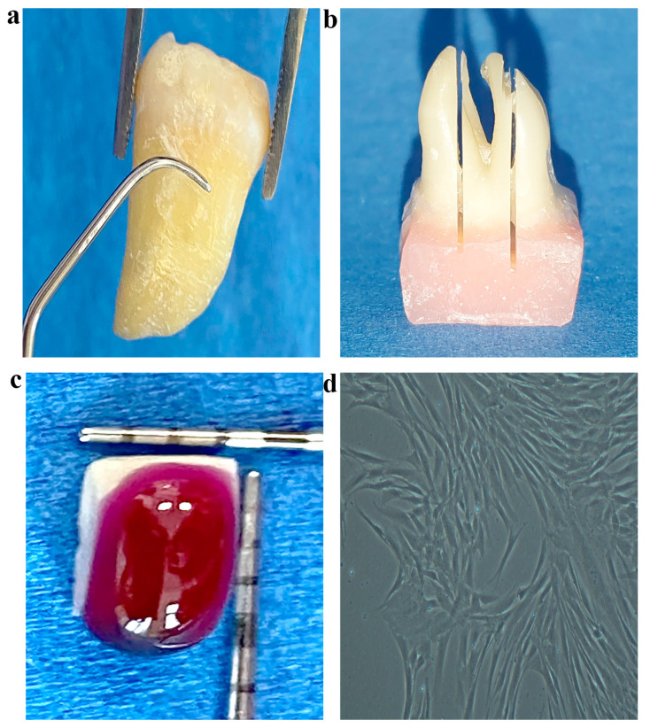
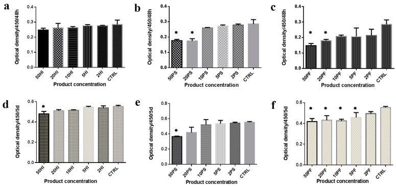
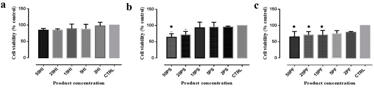
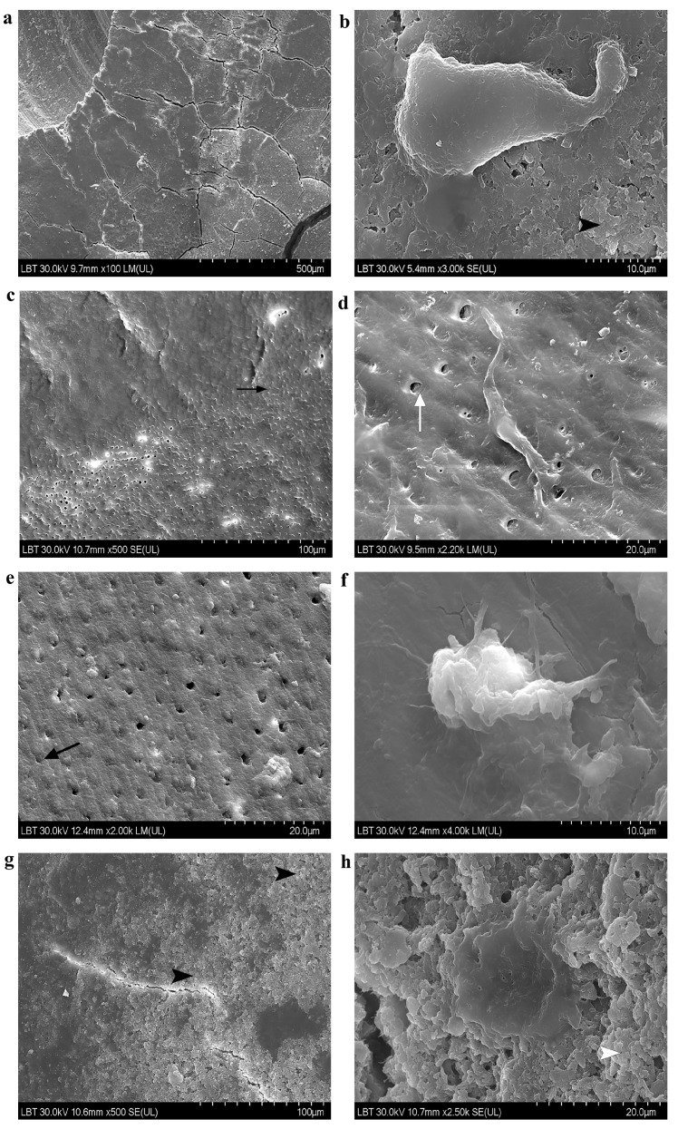
(1) Background: this study aims to test the cytotoxicity of three antimicrobial products used in periodontitis treatment on gingival mesenchymal stem cells (gMSCs) and their influence on root surfaces and gMSC adhesion. We tested the null hypothesis that the effects of the antimicrobials did not differ. (2) Methods: the commercial products based on sulphonic/sulphuric acids, sodium hypochlorite and silver nanoparticles, in five different concentrations, were added to culture medium for growing gMSCs. Cell proliferation capacity was tested using the Cell Counting Kit-8 (CCK8) and their viability was determined by succinate dehydrogenase activity (MTT) assay. Scanning electron microscopy evaluated the adhesion of gMSCs on root samples treated mechanically and with commercial products. (3) Results: the products induced a dose-dependent cytotoxicity in terms of reduced proliferation and viability of gMSCs, as well as cell shape modifications. Significant differences in CCK8 values between the different commercial products were observed. Based on proliferation tests, the null hypothesis was rejected. When MTT values of the three products were compared with each other, no significant differences were observed for any of the five concentrations (p = 0.065, p = 0.067, p = 0.172, p = 0.256, p = 0.060). (4) Conclusions: the three antimicrobials had a certain degree of cytotoxicity on gMSCs. gMSCs repopulated treated root surfaces.

Keywords: adhesion; dental disinfectant; smear layer; stem cell; tooth root.


Conflict of interest statement

The authors declare no conflict of interest.

Figures

Figure 1
Figure 1
Sample preparation for SEM analysis. (a) Root scaling; (b) longitudinally sectioned roots; (c) HI on a standard root sample; (d) MSCs prepared to be harvested at 80% confluence.
Figure 2
Figure 2
Proliferation after 48 h and 5 days of culture in different media. The whiskers correspond to the standard deviation. Results after 48 h for experimental media containing: (a) HI; (b) PS; (c) PF; results after 5 days for experimental media containing (d) HI; (e) PS; (f) PF; HI = HybenX; PS = Perisolv; PF = Perioflush; CTRL = Control. * = statistically different vs. controls.
Figure 3
Figure 3
Graphical representation of MTT test. The whiskers correspond to the standard deviation. Results for experimental media containing: (a) HI; (b) PS; (c) PF; HI = HybenX; PS = Perisolv; PF = Perioflush; CTRL = Control. * = statistically different vs. controls.
Figure 4
Figure 4
SEM observed samples. (a) Continuous smear layer on a instrumented root sample (100×); (b) elongate cell, well-attached through pseudopods on a rough smear-layer covered sample after subgingival instrumentation (3000×); (c) transversally sectioned dentin tubuli partly obstructed by a continuous smooth smear layer (upper part) on a HI-treated and instrumented sample (500×); (d) fusiform well-attached cell through long lamellipodia and dentin tubuli of 3-4 µm on a HI-treated and instrumented sample (2200×); (e) relative smooth smear layer surface partially obstructing dentin tubuli of a PS-treated and instrumented sample (2000×); (f) attached cell through filopodia on a relatively smooth PS-treated and instrumented sample (4000×); (g) rough surface due to important deposits of a PF-treated and instrumented sample (500×); (h) beautiful flattened adherent cell through short lamellipodia on a very rough PF-treated and instrumented sample (2500×). HI = HybenX; PS = Perisolv; PF = Perioflush. Black arrowhead = rough smear-layer; white arrowhead = very rough smear layer; black arrow = partly obstructed dentin tubuli; white arrow = open dentin tubuli.

Similar articles

Cited by

References

    1. Savage A., Eaton K.A., Moles D.R., Needleman I. A systematic review of definitions of periodontitis and methods that have been used to identify this disease. J. Clin. Periodontol. 2009;36:458–467. doi: 10.1111/j.1600-051X.2009.01408.x. - DOI - PubMed
    1. Peres M.A., Macpherson L.M.D., Weyant R.J., Daly B., Venturelli R., Mathur M.R., Listl S., Celeste R.K., Guarnizo-Herreño C.C., Kearns C., et al. Oral diseases: A global public health challenge. Lancet. 2019;394:249–260. doi: 10.1016/S0140-6736(19)31146-8. - DOI - PubMed
    1. Graziani F., Karapetsa D., Alonso B., Herrera D. Nonsurgical and surgical treatment of periodontitis: How many options for one disease? Periodontol. 2000. 2017;75:152–188. doi: 10.1111/prd.12201. - DOI - PubMed
    1. Sanz M., Herrera D., Kebschull M., Chapple I., Jepsen S., Beglundh T., Sculean A., Tonetti M.S., Merete Aass A., Aimetti M., et al. Treatment of stage I–III periodontitis—The EFP S3 level clinical practice guideline. J. Clin. Periodontol. 2020;47:4–60. doi: 10.1111/jcpe.13290. - DOI - PMC - PubMed
    1. Gomes E.W.B., Casarin M., Martins T.M., da Silva A.F. Local delivery therapies as adjuvants to non-surgical periodontal treatment of periodontitis grade C: A systematic review. Clin. Oral Investig. 2000;24:4213–4224. doi: 10.1007/s00784-020-03631-8. - DOI - PubMed

LinkOut - more resources