

The Use of Point-of-Care Ultrasound (POCUS) in the Diagnosis of Deep Vein Thrombosis
- PMID: 34501350
- PMCID: PMC8432124
- DOI: 10.3390/jcm10173903
The Use of Point-of-Care Ultrasound (POCUS) in the Diagnosis of Deep Vein Thrombosis
Abstract
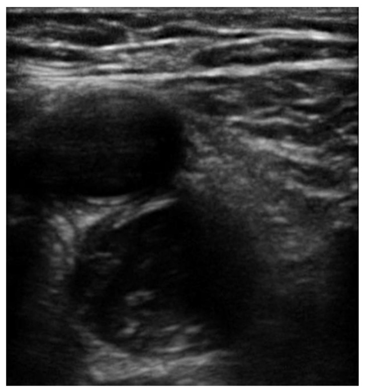
Acute lower extremity proximal deep venous thrombosis (DVT) requires accurate diagnosis and treatment in order to prevent embolization and other complications. Point-of-care ultrasound (POCUS), a clinician performed, and clinician interpreted bedside ultrasound examination has been increasingly used for DVT evaluation mainly in the urgent and critical care setting, but also in the ambulatory clinics and the medical wards. Studies have demonstrated that POCUS has excellent diagnostic accuracy for acute proximal DVT when performed by well-trained users. However, there is significant heterogeneity among studies on the necessary extent of training and universally acceptable standardized education protocols are needed. In this review, we summarize the evidence that supports the use of POCUS to diagnose acute proximal DVT and focus on methodology and current technology, sensitivity and specificity, pre-test probability and the role of D-dimer, time and resources, education, limitations, and future directions.
Keywords: DVT; POCUS; VTE; deep vein thrombosis; point-of-care ultrasound; sonography; ultrasound; venous thromboembolism.
Conflict of interest statement
The authors declare no conflict of interest.
Figures
Similar articles
-
Deep venous thrombosis (DVT) diagnostics: gleaning insights from point-of-care ultrasound (PoCUS) techniques in emergencies: a systematic review and meta-analysis.Ultrasound J. 2024 Jul 30;16(1):37. doi: 10.1186/s13089-024-00378-1. Ultrasound J. 2024. PMID: 39080184 Free PMC article. Review.
-
Point-Of-Care Ultrasound Screening for Proximal Lower Extremity Deep Venous Thrombosis.J Vis Exp. 2023 Feb 10;(192). doi: 10.3791/64601. J Vis Exp. 2023. PMID: 36847361
-
Emergency medicine residents' learning curve in diagnosing deep vein thrombosis with 3-point venous point-of-care ultrasound.Int J Emerg Med. 2024 Jun 17;17(1):75. doi: 10.1186/s12245-024-00645-x. Int J Emerg Med. 2024. PMID: 38886639 Free PMC article.
-
Hospitalist-Operated Compression Ultrasonography: a Point-of-Care Ultrasound Study (HOCUS-POCUS).J Gen Intern Med. 2019 Oct;34(10):2062-2067. doi: 10.1007/s11606-019-05120-5. Epub 2019 Aug 6. J Gen Intern Med. 2019. PMID: 31388904 Free PMC article.
-
Point-of-Care Ultrasound for Bedside Diagnosis of Lower Extremity DVT.Chest. 2021 Nov;160(5):1853-1863. doi: 10.1016/j.chest.2021.07.010. Epub 2021 Jul 13. Chest. 2021. PMID: 34270964 Review.
Cited by
-
Point-of-Care Ultrasound Findings in Occlusive Iliac Vein Thrombus During Pregnancy: A Case Report.Clin Pract Cases Emerg Med. 2024 Aug;8(3):254-258. doi: 10.5811/cpcem.6658. Clin Pract Cases Emerg Med. 2024. PMID: 39158244 Free PMC article.
-
Risk factors and a nomogram model for deep vein thrombosis in critically ill patients with sepsis: a retrospective analysis.Sci Rep. 2025 May 13;15(1):16641. doi: 10.1038/s41598-025-01660-5. Sci Rep. 2025. PMID: 40360645 Free PMC article.
-
How Point-of-Care Ultrasound Led to a Diagnosis of May-Thurner Syndrome.POCUS J. 2021 Nov 23;6(2):76-79. doi: 10.24908/pocus.v6i2.15105. eCollection 2021. POCUS J. 2021. PMID: 36895671 Free PMC article.
-
Venous Thromboembolism in Pregnancy: Challenges and Solutions.Vasc Health Risk Manag. 2023 Jul 20;19:469-484. doi: 10.2147/VHRM.S404537. eCollection 2023. Vasc Health Risk Manag. 2023. PMID: 37492280 Free PMC article. Review.
-
The incidence and risk factors of proximal lower extremity deep vein thrombosis without pharmacologic prophylaxis in critically ill surgical Taiwanese patients: A prospective study.J Intensive Care Soc. 2023 Dec 28;25(2):140-146. doi: 10.1177/17511437231214906. eCollection 2024 May. J Intensive Care Soc. 2023. PMID: 38737310 Free PMC article.
References
-
- Pomero F., Dentali F., Borretta V., Bonzini M., Melchio R., Douketis J.D., Fenoglioet L.M. Accuracy of emergency physician-performed ultrasonography in the diagnosis of deep-vein thrombosis: A systematic review and meta-analysis. Thromb. Haemost. 2013;109:137–145. doi: 10.1160/TH12-07-0473. - DOI - PubMed
Publication types
LinkOut - more resources
Full Text Sources
