

Interstitial Lung Disease at High Resolution CT after SARS-CoV-2-Related Acute Respiratory Distress Syndrome According to Pulmonary Segmental Anatomy
- PMID: 34501430
- PMCID: PMC8432464
- DOI: 10.3390/jcm10173985
Interstitial Lung Disease at High Resolution CT after SARS-CoV-2-Related Acute Respiratory Distress Syndrome According to Pulmonary Segmental Anatomy
Abstract
Background: The purpose of this study was to evaluate High-Resolution CT (HRCT) findings in SARS-CoV-2-related ARDS survivors treated with prolonged low-dose methylprednisolone after hospital discharge.
Methods: A total of 44 consecutive patients (M: 32, F: 12, average age: 64), hospitalised in our department from April to September 2020 for SARS-CoV-2-related ARDS, who had a postdischarge CT scan, were enrolled into this retrospective study. We reviewed the electronic medical charts to collect laboratory, clinical, and demographic data. The CT findings were evaluated and classified according to lung segmental distribution. The imaging findings were correlated with spirometry results and included ground glass opacities (GGOs), consolidations, reticulations, bronchiectasis/bronchiolectasis, linear bands, and loss of pulmonary volume.
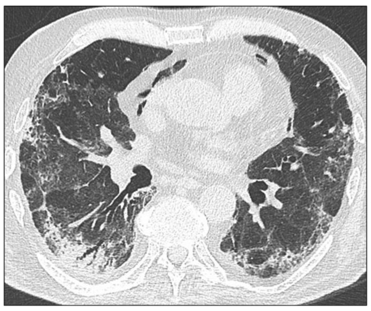
Results: Alterations in the pulmonary parenchyma were observed in 97.7% of patients at HRCT (median time lapse between ARDS diagnosis and HRCT: 2.8 months, range 0.9 to 6.7). The most common findings were linear bands (84%), followed by GGOs (75%), reticulations (34%), bronchiolectasis (32%), consolidations (30%), bronchiectasis (30%) and volume loss (25%). They had a symmetric distribution, and both lower lobes were the most affected areas.
Conclusions: A reticular pattern with a posterior distribution was observed 3 months after discharge from severe COVID-19 pneumonia, and this differs from previously described postCOVID-19 fibrotic-like changes. We hypothesized that the systematic use of prolonged low-dose of corticosteroid could be the main reason of this different CT scan appearance.
Keywords: COVID-19 pneumonia; acute respiratory distress syndrome; high resolution computed tomography; pulmonary fibrosis.
Conflict of interest statement
The authors declare no conflict of interest.
Figures


References
-
- The WHO Rapid Evidence Appraisal for COVID-19 Therapies (REACT) Working Group. Sterne J., Murthy S., Diaz J.V., Slutsky A.S., Villar J., Angus D.C., Annane D., Azevedo L.C.P., Berwanger O., et al. Association Between Administration of Systemic Corticosteroids and Mortality Among Critically Ill Patients With COVID-19: A Meta-analysis. JAMA. 2020;324:1330–1341. doi: 10.1001/jama.2020.17023. - DOI - PMC - PubMed
LinkOut - more resources
Full Text Sources
Miscellaneous
