Review
. 2021 Sep 3;10(17):3993.
doi: 10.3390/jcm10173993.

Peripheral Manifestations in Age Related Macular Degeneration: A Review of Imaging and Findings

Affiliations
Review

Peripheral Manifestations in Age Related Macular Degeneration: A Review of Imaging and Findings

Andrew Pivovar et al. J Clin Med. .

Abstract

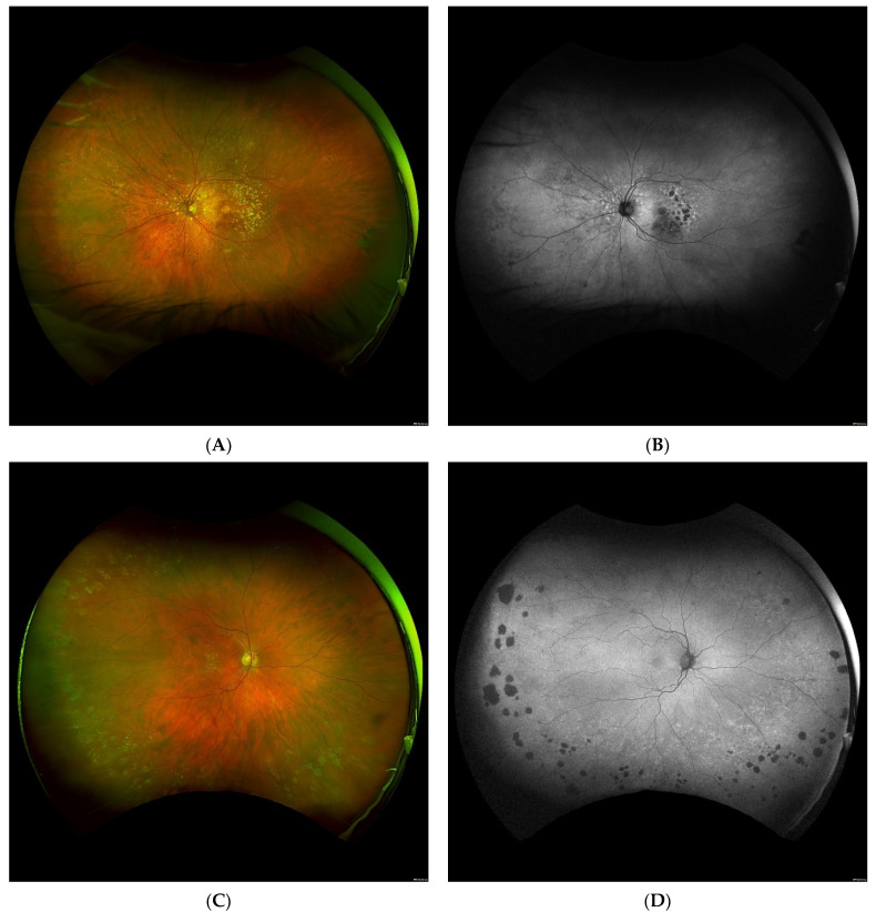
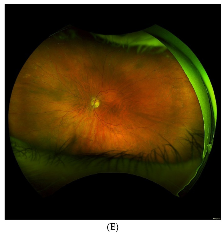
Purpose: To review novel findings in research with ultra-widefield imaging for analysis of peripheral manifestations in macular degeneration (AMD). We introduce the evolving widefield imaging modalities while summarizing the analytical techniques used in data collection of peripheral retinal findings thus far. Our review provides a summary of advancements to date and a commentary on future direction for AMD research.

Methods: This is a literature review of all significant publications focused on the relationship between AMD and the retinal periphery conducted within the last two decades.

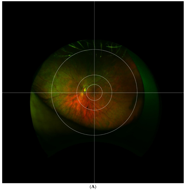
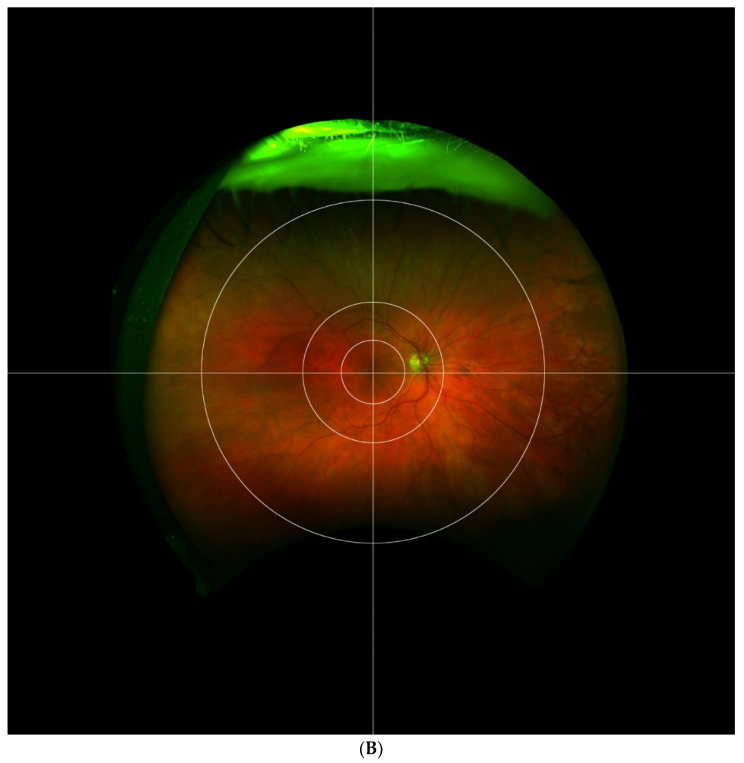
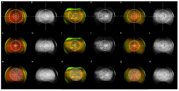
Results and conclusion: Promising research has been undertaken to elucidate peripheral retinal manifestations in macular degeneration using novel methodology. Advancements in ultra-widefield imaging and fundus autofluorescence have allowed us to elucidate peripheral retinal pigmentary changes, drusen deposition, and much more. Novel grid overlay techniques have been introduced to aid in analyzing these changes for pattern recognition and grouping of findings. This review discusses these findings in detail, providing evidence for the pan-retinal manifestations of AMD. Inter-study discordance in analytical approach highlights a need for more systematic future study.

Keywords: age-related macular degeneration; fluorescein autofluorescence; grid analysis; peripheral; ultra-widefield.


Conflict of interest statement

The authors declare no conflict of interest.

Figures

Figure 1
Figure 1
Exemplary ultra-widefield images of eyes with macular degeneration and peripheral abnormalities. Pseudocolor and corresponding autofluorescence of a left eye demonstrating multifocal retinal pigment epithelium (RPE) atrophy and drusen in the macula with peripheral drusen and multifocal RPE atrophy, especially nasal to the optic nerve. Note there is a glaucomatous disk hemorrhage and a peripheral retinal tear status post laser retinopexy (A,B). Another example demonstrates a right eye with macular drusen and peripheral drusen and multifocal atrophy in the far periphery (C,D). A left eye with macular drusen and pigment changes and drusenoid bodies and mild reticular pseudopigmentation in the periphery. Note eyelash artifact inferior (E).
Figure 1
Figure 1
Exemplary ultra-widefield images of eyes with macular degeneration and peripheral abnormalities. Pseudocolor and corresponding autofluorescence of a left eye demonstrating multifocal retinal pigment epithelium (RPE) atrophy and drusen in the macula with peripheral drusen and multifocal RPE atrophy, especially nasal to the optic nerve. Note there is a glaucomatous disk hemorrhage and a peripheral retinal tear status post laser retinopexy (A,B). Another example demonstrates a right eye with macular drusen and peripheral drusen and multifocal atrophy in the far periphery (C,D). A left eye with macular drusen and pigment changes and drusenoid bodies and mild reticular pseudopigmentation in the periphery. Note eyelash artifact inferior (E).
Figure 2
Figure 2
The Boston grid is depicted superimposed on fundus images of right (A) and left (B) eyes. The grid is composed of 3 concentric circles centered on the fovea, separating the retina into central, perimacular, mid-peripheral, and far-peripheral areas. Horizontal and vertical lines further separate the retina into superior, inferior, temporal, and nasal quadrants, producing a total of 12 zones.
Figure 2
Figure 2
The Boston grid is depicted superimposed on fundus images of right (A) and left (B) eyes. The grid is composed of 3 concentric circles centered on the fovea, separating the retina into central, perimacular, mid-peripheral, and far-peripheral areas. Horizontal and vertical lines further separate the retina into superior, inferior, temporal, and nasal quadrants, producing a total of 12 zones.
Figure 3
Figure 3
A comparison of the Boston grid to two other discussed grids for a total of 3 patients with AMD (AF). Fundus pseudocolor and autofluorescence imaging of 3 patients diagnosed with AMD with the Boston grid overlay. Fundus imaging of the first patient is depicted in (A,B,G,H,M,N). Peri-macular drusen in (C) is best captured by the Boston grid when compared with the Lengyel (I) and Reznicek (O) grids. Similarly, central drusen in (E) is best circumscribed by the Boston grid. (GL) The exact same photos as in (AF) with the recreated grid by Lengyel et al. superimposed. In comparison to the Boston grid, the Lengyel grid lacks comprehensive peri-macular and mid-peripheral zones, with zones 1–3 found strictly in the macula and zone 4 encompassing both the mid-periphery and peri-macular area. (MR) An overlay of the recreated Reznicek et al. grid on the images presented previously. The 48 partitions seen here do not clearly distinguish the mid-periphery from the far-periphery and peri-macular areas of the retina, but create much further detail which might be helpful in select cases. For instance, the peri-macular drusen in (O) can be found in approximately 12 zones as opposed to one or few distinct zones. Grids were recreated based on instructions detailed elsewhere [13,20].

References

    1. Gheorghe A., Mahdi L., Musat O. Age-Related Macular Degeneration. Rom. J. Ophthalmol. 2015;59:74–77. - PMC - PubMed
    1. Oellers P., Laíns I., Mach S., Garas S., Kim I.K., Vavvas D.G., Miller J.W., Husain D., Miller J.B. Novel grid combined with peripheral distortion correction for ultra-widefield image grading of age-related macular degeneration. Clin. Ophthalmol. 2017;11:1967–1974. doi: 10.2147/OPTH.S143246. - DOI - PMC - PubMed
    1. Patel S.N., Shi A., Wibbelsman T.D., Klufas M.A. Ultra-widefield retinal imaging: An update on recent advances. Ther. Adv. Ophthalmol. 2020;12:2515841419899495. doi: 10.1177/2515841419899495. - DOI - PMC - PubMed
    1. Choudhry N., Duker J.S., Freund K.B., Kiss S., Querques G., Rosen R., Sarraf D., Souied E.H., Stanga P.E., Staurenghi G., et al. Classification and Guidelines for Widefield Imaging: Recommendations from the International Widefield Imaging Study Group. Ophthalmol. Retin. 2019;3:843–849. doi: 10.1016/j.oret.2019.05.007. - DOI - PubMed
    1. Nagiel A., Lalane R.A., Sadda S.R., Schwartz S.D. Ultra-Widefield Fundus Imaging. Retina. 2016;36:660–678. doi: 10.1097/IAE.0000000000000937. - DOI - PubMed

LinkOut - more resources