. 2021 Aug 24;22(17):9124.
doi: 10.3390/ijms22179124.

Exploring the Binding Mechanism of PF-07321332 SARS-CoV-2 Protease Inhibitor through Molecular Dynamics and Binding Free Energy Simulations

Affiliations

Exploring the Binding Mechanism of PF-07321332 SARS-CoV-2 Protease Inhibitor through Molecular Dynamics and Binding Free Energy Simulations

Bilal Ahmad et al. Int J Mol Sci. .

Abstract

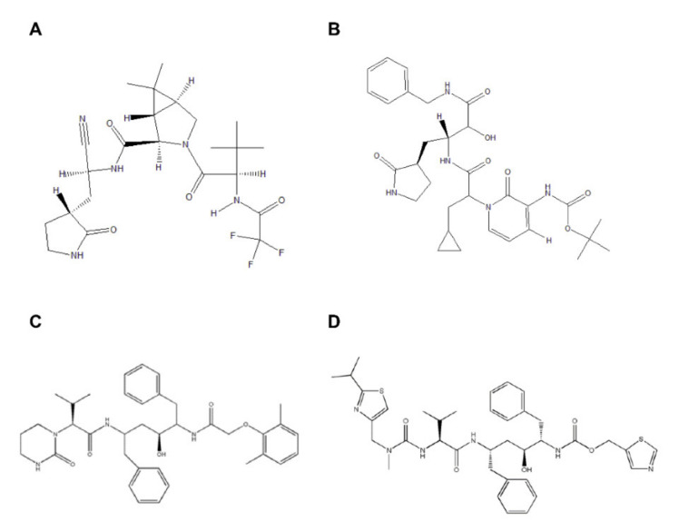
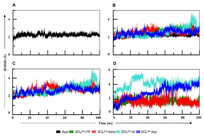
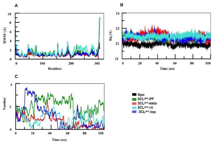
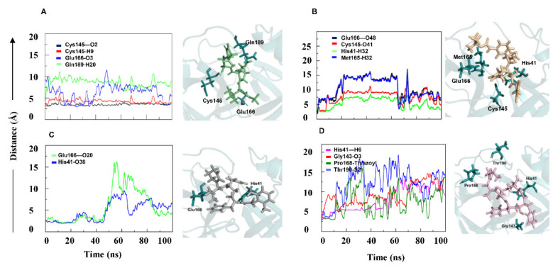
The novel coronavirus disease, caused by severe acute respiratory coronavirus 2 (SARS-CoV-2), rapidly spreading around the world, poses a major threat to the global public health. Herein, we demonstrated the binding mechanism of PF-07321332, α-ketoamide, lopinavir, and ritonavir to the coronavirus 3-chymotrypsin-like-protease (3CLpro) by means of docking and molecular dynamic (MD) simulations. The analysis of MD trajectories of 3CLpro with PF-07321332, α-ketoamide, lopinavir, and ritonavir revealed that 3CLpro-PF-07321332 and 3CLpro-α-ketoamide complexes remained stable compared with 3CLpro-ritonavir and 3CLpro-lopinavir. Investigating the dynamic behavior of ligand-protein interaction, ligands PF-07321332 and α-ketoamide showed stronger bonding via making interactions with catalytic dyad residues His41-Cys145 of 3CLpro. Lopinavir and ritonavir were unable to disrupt the catalytic dyad, as illustrated by increased bond length during the MD simulation. To decipher the ligand binding mode and affinity, ligand interactions with SARS-CoV-2 proteases and binding energy were calculated. The binding energy of the bespoke antiviral PF-07321332 clinical candidate was two times higher than that of α-ketoamide and three times than that of lopinavir and ritonavir. Our study elucidated in detail the binding mechanism of the potent PF-07321332 to 3CLpro along with the low potency of lopinavir and ritonavir due to weak binding affinity demonstrated by the binding energy data. This study will be helpful for the development and optimization of more specific compounds to combat coronavirus disease.

Keywords: 3CL protease; COVID-19; PF-07321332; SARS-CoV-2; main protease; α-ketoamide.


Conflict of interest statement

The authors declare no conflict of interest.

Figures

Figure 1
Figure 1
2D structure representation of (A) PF-07321332, (B) α-ketoamide, (C) lopinavir, and (D) ritonavir.
Figure 2
Figure 2
(A) The root mean square deviation (RMSD) graph of the apo form of 3CLpro during the 100 ns simulation. The graph suggests that protein was stable during the whole simulation, with an average RMSD of 2.2 Å. (B) The RMSD graphs of all four proteins in complexes in comparison with the apo form. The protein in 3CLpro–PF and 3CLpro–keto remained stable whereas in 3CLpro–lop and 3CLpro–rit, it showed more fluctuations than in the other two 3CLpro systems. (C) The RMSD graph of 3CLpro complexes. The 3CLpro–rit and 3CLpro–lop complexes showed similar trend as the proteins. (D) RMSD graphs of the ligands in complexes with 3CLpro. The ligands PF-07321332 and α-ketoamide seemed stable, whereas both lopinavir and ritonavir in 3CLpro complexes featured noticeable fluctuations. PF—PF-07321332; keto—α-ketoamide; rit—ritonavir; lop—lopinavir.
Figure 3
Figure 3
(A) Root mean square fluctuation graphs of apo-3CLpro and of 3CLpro complexes. In comparison with the apo-form, regions spanning amino acid residues 45–75 and 150–200 in both docked complexes showed deviations. (B) Radius of gyration of the 3CLpro in complexes was higher than that of its apo-form. (C) Number of hydrogen bonds in the four complexes. PF—PF-07321332; keto—α-ketoamide; rit—ritonavir; lop—lopinavir.
Figure 4
Figure 4
The minimum distance graph of protein–ligand interactions. The interaction distance between the protein and ligand in 3CLpro–PF (A) and 3CLpro–keto (B) remained constant. Interaction in 3CLpro–lop (C) initially remained constant but showed major fluctuations afterwards. In 3CLpro–rit (D), the lengths of all four bonds continuously increased. PF—PF-07321332; keto—α-ketoamide; rit—ritonavir; lop—lopinavir.

References

    1. Velavan T.P., Meyer C.G. The COVID-19 epidemic. Trop. Med. Int. Health. 2020;25:278–280. doi: 10.1111/tmi.13383. - DOI - PMC - PubMed
    1. Johns Hopkins Coronavirus Resource Center COVID-19 Map. [(accessed on 10 August 2020)]; Available online: https://coronavirus.jhu.edu/map.html.
    1. Dai W., Zhang B., Su H., Li J., Zhao Y., Xie X., Jin Z., Liu F., Li C., Li Y., et al. Structure-based design of antiviral drug candidates targeting the SARS-CoV-2 main protease. Science. 2020;368:1331–1335. doi: 10.1126/science.abb4489. - DOI - PMC - PubMed
    1. Zhou P., Yang X.L., Wang X.G., Hu B., Zhang L., Zhang W., Si H.R., Zhu Y., Li B., Huang C.L., et al. A pneumonia outbreak associated with a new coronavirus of probable bat origin. Nature. 2020;579:270–273. doi: 10.1038/s41586-020-2012-7. - DOI - PMC - PubMed
    1. Lu R., Zhao X., Li J., Niu P., Yang B., Wu H., Wang W., Song H., Huang B., Zhu N., et al. Genomic characterisation and epidemiology of 2019 novel coronavirus: Implications for virus origins and receptor binding. Lancet. 2020;395:565–574. doi: 10.1016/S0140-6736(20)30251-8. - DOI - PMC - PubMed

MeSH terms

LinkOut - more resources