. 2021 Aug 24;22(17):9125.
doi: 10.3390/ijms22179125.

Enhancement of Immune Checkpoint Inhibitor-Mediated Anti-Cancer Immunity by Intranasal Treatment of Ecklonia cava Fucoidan against Metastatic Lung Cancer

Affiliations

Enhancement of Immune Checkpoint Inhibitor-Mediated Anti-Cancer Immunity by Intranasal Treatment of Ecklonia cava Fucoidan against Metastatic Lung Cancer

Wei Zhang et al. Int J Mol Sci. .

Abstract

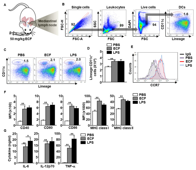
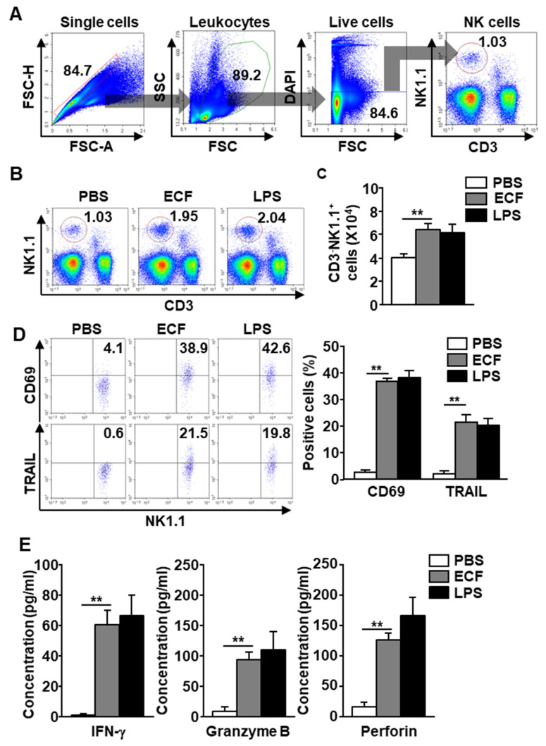
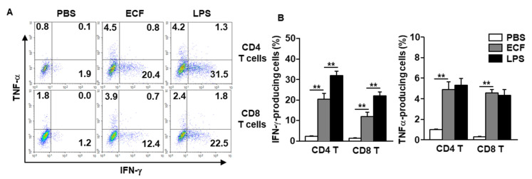
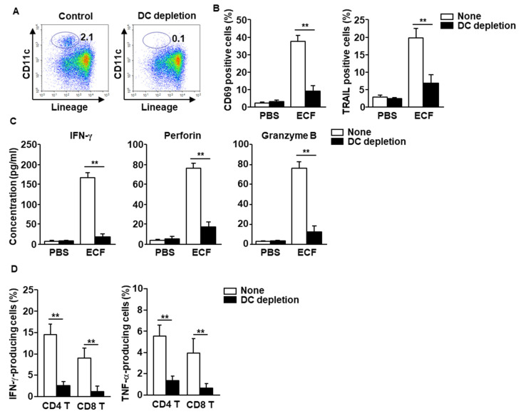
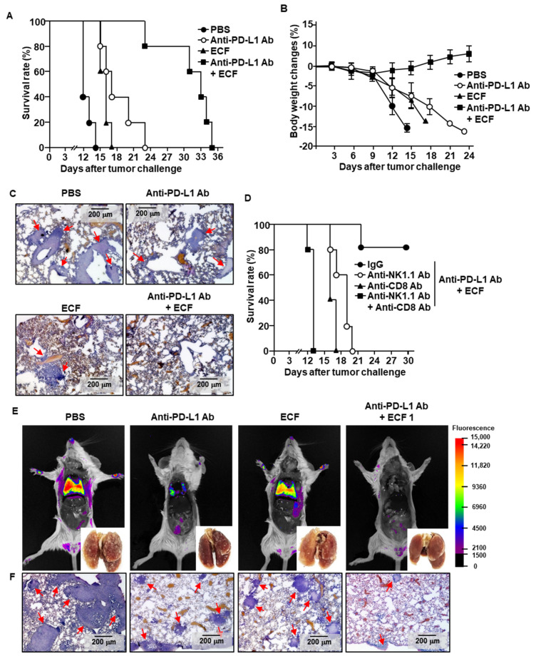
Although fucoidan, a well-studied seaweed-extracted polysaccharide, has shown immune stimulatory effects that elicit anticancer immunity, mucosal adjuvant effects via intranasal administration have not been studied. In this study, the effect of Ecklonia cava-extracted fucoidan (ECF) on the induction of anti-cancer immunity in the lung was examined by intranasal administration. In C57BL/6 and BALB/c mice, intranasal administration of ECF promoted the activation of dendritic cells (DCs), natural killer (NK) cells, and T cells in the mediastinal lymph node (mLN). The ECF-induced NK and T cell activation was mediated by DCs. In addition, intranasal injection with ECF enhanced the anti-PD-L1 antibody-mediated anti-cancer activities against B16 melanoma and CT-26 carcinoma tumor growth in the lungs, which were required cytotoxic T lymphocytes and NK cells. Thus, these data demonstrated that ECF functioned as a mucosal adjuvant that enhanced the immunotherapeutic effect of immune checkpoint inhibitors against metastatic lung cancer.

Keywords: Ecklonia cava fucoidan; anti-PD-L1 antibody; anti-cancer; immunotherapy; mucosal adjuvant.


Conflict of interest statement

The authors declare no conflict of interest.

Figures

Figure 1
Figure 1
Dendritic cell (DC) activation in the mediastinal lymph node (mLN) by intranasal administration of Ecklonia cava fucoidan (ECF). (A) C57BL/6 mice were intranasally (i.n.) administered with 50 mg/kg ECF. The DC activation in mLN was harvested and analyzed 18 h after ECF administration. (B) The definition of DCs in mLNs was shown. (C) Percentage of mLN DCs, as analyzed by flow cytometry. PBS, phosphate-buffered saline; LPS, lipopolysaccharide. (D) Absolute number of DCs in the mLN. (n = 6, two-way analysis of variance, ** p < 0.01) (E) Expression levels of C-C chemokine receptor type 7 (CCR7) in mLN DCs were shown. IgG, immunoglobulin G. (F) Costimulatory molecules and major histocompatibility complex (MHC) class I and II expression levels were measured in mLN DCs. MFI, mean fluorescence intensity; (G) The levels of interleukin (IL)-6, IL-12p40, and tumor necrosis factor (TNF)-α levels in bronchoalveolar lavage (BAL) fluid. (n = 6, two-way analysis of variance, * p < 0.05 and ** p < 0.01).
Figure 2
Figure 2
Natural killer (NK) cell activation by ECF. Twenty-four hours after 50 mg/kg ECF administration intranasally, the mLN was harvested from C57BL/6 mice. (A) NK cells definition was shown. (B) The percentage of NK cells was shown. PBS, phosphate-buffered saline; LPS, lipopolysaccharide. (C) Average of NK cell number in the mLN (n = 6, two-way analysis of variance, ** p < 0.01). (D) Surface activation markers, CD69 and tumor necrosis factor-related apoptosis-inducing ligand (TRAIL) expression levels in mLN NK cells were shown (left panel). Mean number of CD69 and TRAIL positive cells in mLN NK cells were shown (right panel, n = 6, two-way analysis of variance, ** p < 0.01). (E) Concentration of interferon (IFN)-γ, granzyme B, and perforin levels in BAL fluid was shown. (n = 6, two-way analysis of variance, ** p < 0.01).
Figure 3
Figure 3
Induction of T helper 1 and cytotoxic T 1 (Tc1) immune responses in the mLN by intranasal administration with ECF. C57BL/6 mice were i.n. administered with 50 mg/kg ECF twice at a 3-day interval. (A) Intracellular producing levels of IFN-γ and TNF-α in mLN CD4 and CD8 T cells were measured. (B) Mean percentage of IFN-γ-producing (left panel) and TNF-α-producing (right panel) CD4 and CD8 T cells (n = 6, two-way analysis of variance, ** p < 0.01).
Figure 4
Figure 4
DCs are required for stimulation of NK and T cells in the mLN. (A) DCs in mLN cells were depleted, as described in the Methods. (B) CD69 and tumor necrosis factor-related apoptosis-inducing ligand (TRAIL) expression levels, as measured 24 h after treatment with 100 μg/mL of ECF. PBS, phosphate-buffered saline (n = 6, two-way analysis of variance, ** p < 0.01). (C) The concentration of IFN-γ, perforin, and granzyme B in the cultured medium was shown. (D) Percentage of intracellular IFN-γ- and TNF-α-producing T cells in mLN cells after culture with or without 100 μg/mL ECF for 3 days, as analyzed by flow cytometry (n = 6, two-way analysis of variance).
Figure 5
Figure 5
ECF enhances anti-PD-L1 antibody-induced anti-cancer immunity. C57BL/6 mice were challenged intravenously (i.v.) with 0.5 × 10⁶ B16 cells. As indicated in the materials and method, the mice were treated with PBS 10 mg/kg of anti-PD-L1 antibody (Ab), 50 mg/kg of ECF and the combination of ECF and anti-PD-L1 Ab. (A) The graph showed survival rate of treated mice (n = 5 for each group). (B) Changes of body weight during treatment were shown. (C) Hematoxylin and eosin (H&E) staining of lung showed B16 melanoma cell infiltration, as analyzed on day 10 after tumor injection. Red arrows indicated infiltrated tumor cells. (D) Survival rate, as measured in natural killer (NK)- or CD8 T cell-depleted mice (n = 5 for each group). (E,F) BALB/c mice were injected i.v. with 0.5 × 10⁶ CT-26-iRFP cells. The mice received with PBS, 10 mg/kg of anti-PD-L1 Ab, 50 mg/kg of ECF or the combination of anti-PD-L1 Ab and ECF for a 3 day-interval. (E) Fluorescence imaging of iRFP in the mice on day 14 after CT-26-iRFP cell administration in BLAB/c mice, n = 5. (F) CT-26 cell infiltration in lung was analyzed by H&E staining of lung tissue 14 days after CT-26 injection in BALB/c mice. Red arrows indicated infiltrated tumor cells.

References

    1. Patel S. Therapeutic importance of sulfated polysaccharides from seaweeds: Updating the recent findings. 3 Biotech. 2012;2:171–185. doi: 10.1007/s13205-012-0061-9. - DOI
    1. Manlusoc J.K.T., Hsieh C.-L., Hsieh C.-Y., Salac E.S.N., Lee Y.-T., Tsai P.-W. Pharmacologic application potentials of sulfated polysaccharide from marine algae. Polymers. 2019;11:1163. doi: 10.3390/polym11071163. - DOI - PMC - PubMed
    1. Kidgell J.T., Magnusson M., de Nys R., Glasson C.R. Ulvan: A systematic review of extraction, composition and function. Algal Res. 2019;39:101422. doi: 10.1016/j.algal.2019.101422. - DOI
    1. Jin J.-O., Chauhan P.S., Arukha A.P., Chavda V., Dubey A., Yadav D. The Therapeutic Potential of the Anticancer Activity of Fucoidan: Current Advances and Hurdles. Mar. Drugs. 2021;19:265. doi: 10.3390/md19050265. - DOI - PMC - PubMed
    1. Zhang Y.-Q., Tsai Y.-C., Monie A., Hung C.-F., Wu T.-C. Carrageenan as an adjuvant to enhance peptide-based vaccine potency. Vaccine. 2010;28:5212–5219. doi: 10.1016/j.vaccine.2010.05.068. - DOI - PMC - PubMed

MeSH terms