

Regulation of Oxygen Homeostasis at the Intestinal Epithelial Barrier Site
- PMID: 34502078
- PMCID: PMC8431628
- DOI: 10.3390/ijms22179170
Regulation of Oxygen Homeostasis at the Intestinal Epithelial Barrier Site
Abstract
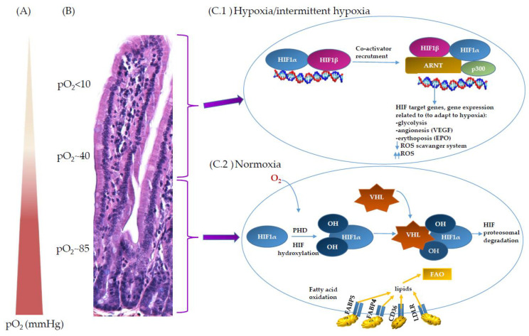
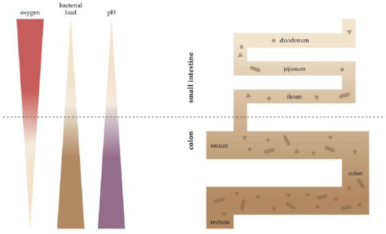
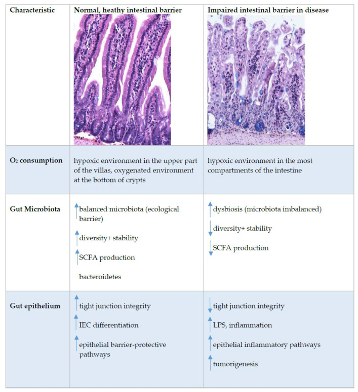
The unique biology of the intestinal epithelial barrier is linked to a low baseline oxygen pressure (pO2), characterised by a high rate of metabolites circulating through the intestinal blood and the presence of a steep oxygen gradient across the epithelial surface. These characteristics require tight regulation of oxygen homeostasis, achieved in part by hypoxia-inducible factor (HIF)-dependent signalling. Furthermore, intestinal epithelial cells (IEC) possess metabolic identities that are reflected in changes in mitochondrial function. In recent years, it has become widely accepted that oxygen metabolism is key to homeostasis at the mucosae. In addition, the gut has a vast and diverse microbial population, the microbiota. Microbiome-gut communication represents a dynamic exchange of mediators produced by bacterial and intestinal metabolism. The microbiome contributes to the maintenance of the hypoxic environment, which is critical for nutrient absorption, intestinal barrier function, and innate and/or adaptive immune responses in the gastrointestinal tract. In this review, we focus on oxygen homeostasis at the epithelial barrier site, how it is regulated by hypoxia and the microbiome, and how oxygen homeostasis at the epithelium is regulated in health and disease.
Keywords: IBD; hypoxia; microbiota; mitochondria; oxygen.
Conflict of interest statement
The authors declare no conflict of interest.
Figures
Similar articles
-
Oxygen battle in the gut: Hypoxia and hypoxia-inducible factors in metabolic and inflammatory responses in the intestine.J Biol Chem. 2020 Jul 24;295(30):10493-10505. doi: 10.1074/jbc.REV120.011188. Epub 2020 Jun 5. J Biol Chem. 2020. PMID: 32503843 Free PMC article. Review.
-
Impact of Bacterial Metabolites on Gut Barrier Function and Host Immunity: A Focus on Bacterial Metabolism and Its Relevance for Intestinal Inflammation.Front Immunol. 2021 May 26;12:658354. doi: 10.3389/fimmu.2021.658354. eCollection 2021. Front Immunol. 2021. PMID: 34122415 Free PMC article. Review.
-
Development, validation and implementation of an in vitro model for the study of metabolic and immune function in normal and inflamed human colonic epithelium.Dan Med J. 2015 Jan;62(1):B4973. Dan Med J. 2015. PMID: 25557335 Review.
-
The Impact of Dietary Sphingolipids on Intestinal Microbiota and Gastrointestinal Immune Homeostasis.Front Immunol. 2021 May 14;12:635704. doi: 10.3389/fimmu.2021.635704. eCollection 2021. Front Immunol. 2021. PMID: 34054805 Free PMC article. Review.
-
Hypoxia inducible factor-1α-induced interleukin-33 expression in intestinal epithelia contributes to mucosal homeostasis in inflammatory bowel disease.Clin Exp Immunol. 2017 Mar;187(3):428-440. doi: 10.1111/cei.12896. Epub 2016 Dec 6. Clin Exp Immunol. 2017. PMID: 27921309 Free PMC article.
Cited by
-
From the Dish to the Real World: Modeling Interactions between the Gut and Microorganisms in Gut Organoids by Tailoring the Gut Milieu.Int J Stem Cells. 2022 Feb 28;15(1):70-84. doi: 10.15283/ijsc21243. Int J Stem Cells. 2022. PMID: 35220293 Free PMC article. Review.
-
Hypoxia and Intestinal Inflammation: Common Molecular Mechanisms and Signaling Pathways.Int J Mol Sci. 2023 Jan 26;24(3):2425. doi: 10.3390/ijms24032425. Int J Mol Sci. 2023. PMID: 36768744 Free PMC article. Review.
-
Metabolic requirements of Th17 cells and of B cells: Regulation and defects in health and in inflammatory diseases.Front Immunol. 2022 Oct 14;13:990794. doi: 10.3389/fimmu.2022.990794. eCollection 2022. Front Immunol. 2022. PMID: 36311757 Free PMC article. Review.
-
Antimicrobial Properties and Mode of Action of Cryptdin-4, a Mouse α-Defensin Regulated by Peptide Redox Structures and Bacterial Cultivation Conditions.Antibiotics (Basel). 2023 Jun 14;12(6):1047. doi: 10.3390/antibiotics12061047. Antibiotics (Basel). 2023. PMID: 37370366 Free PMC article.
-
Skin immunity: dissecting the complex biology of our body's outer barrier.Mucosal Immunol. 2022 Apr;15(4):551-561. doi: 10.1038/s41385-022-00505-y. Epub 2022 Mar 31. Mucosal Immunol. 2022. PMID: 35361906 Review.
References
Publication types
MeSH terms
Substances
LinkOut - more resources
Full Text Sources
