Review
. 2021 Aug 26;22(17):9207.
doi: 10.3390/ijms22179207.

Tau Protein Interaction Partners and Their Roles in Alzheimer's Disease and Other Tauopathies

Affiliations
Review

Tau Protein Interaction Partners and Their Roles in Alzheimer's Disease and Other Tauopathies

Jakub Sinsky et al. Int J Mol Sci. .

Abstract

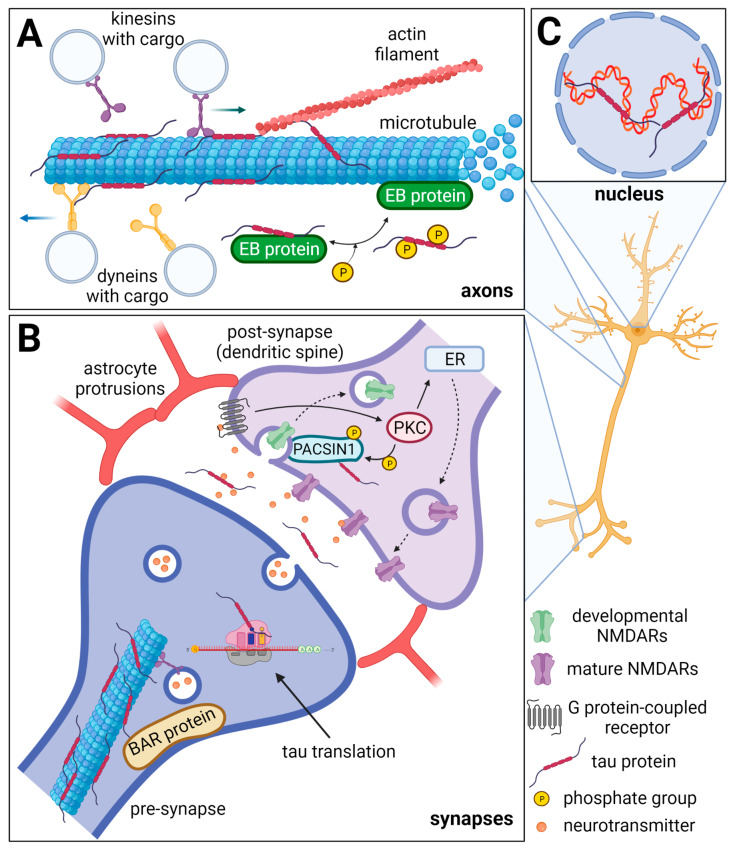
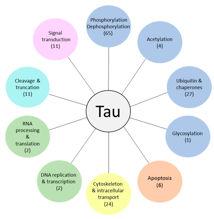
Tau protein plays a critical role in the assembly, stabilization, and modulation of microtubules, which are important for the normal function of neurons and the brain. In diseased conditions, several pathological modifications of tau protein manifest. These changes lead to tau protein aggregation and the formation of paired helical filaments (PHF) and neurofibrillary tangles (NFT), which are common hallmarks of Alzheimer's disease and other tauopathies. The accumulation of PHFs and NFTs results in impairment of physiological functions, apoptosis, and neuronal loss, which is reflected as cognitive impairment, and in the late stages of the disease, leads to death. The causes of this pathological transformation of tau protein haven't been fully understood yet. In both physiological and pathological conditions, tau interacts with several proteins which maintain their proper function or can participate in their pathological modifications. Interaction partners of tau protein and associated molecular pathways can either initiate and drive the tau pathology or can act neuroprotective, by reducing pathological tau proteins or inflammation. In this review, we focus on the tau as a multifunctional protein and its known interacting partners active in regulations of different processes and the roles of these proteins in Alzheimer's disease and tauopathies.

Keywords: Alzheimer’s disease; interaction partners; tau protein; tauopathies.


Conflict of interest statement

The authors declare no conflict of interest.

Figures

Figure 1
Figure 1
Schematic representation of physiological tau protein functions in neurons. The axonal tau (A) stabilizes microtubules (MTs), and it can also bind actin filaments thus facilitating cytoskeleton networking. Furthermore, tau regulates MT dynamics by interacting with end-binding (EB) proteins. The EB proteins promote and regulate MT nucleation and elongation. Tau inhibits the EB protein binding to MTs and this inhibition is reversed by tau phosphorylation. Tau also competitively inhibits the interaction of dynein and kinesin to MTs and thus influences the intraneuronal transport and cargo release. In the synapses (B), tau protein can be directly translated, and during neuronal activity, it is released into the synaptic cleft. Through linking the MTs and actin filaments, tau can influence synaptic plasticity. Moreover, tau is a known interacting partner of the BAR domain-containing proteins such as BAIAP2, PACSIN1, and BIN1, that ensure the curvature and shaping of the neuronal membrane. Tau may play a role in the removal of developmental NMDARs and their replacement for mature NMDARs in dendrites (dashed arrows) as it is a known substrate of the protein kinase C (PKC), which is activated by the G protein-coupled receptors (GPCR). The PACSIN1 recruits clathrin and dynamin endocytic machinery to the developmental NMDARs and thus mediates their removal. The developmental/mature NMDARs exchange is important for the formation of new synaptic connections. ER—endoplasmic reticulum. In the nucleus (C), tau interacts with DNA and RNA, maintains their integrity, and protects them from oxidative damage. Furthermore, tau may be involved in rRNA-coding DNA transcription and rRNA processing. Created with BioRender.com.
Figure 2
Figure 2
Schematic representation of six human tau protein isoforms and their domains. The N-terminal part is more acidic due to N1 and N2 inserts. The microtubule-binding domain (MTBD) is composed of repeats R1–R4. The number of repeats and N-terminal inserts varies according to the type of tau protein isoform.
Figure 3
Figure 3
Schematic representation of human Big tau. Its counterparts in rat and mouse possess 752 or 733 aa and share 74.9% or 74.2% identity, respectively. Exons 4a and 6 are responsible for differentiation from the brain tau protein.
Figure 4
Figure 4
Overview of validated TIPs distributed according to their function. In brackets: the number of validated TIPs for the particular group.

References

    1. Uversky V.N. Intrinsic disorder, protein–protein interactions, and disease. Adv. Protein Chem. Struct. Biol. 2018;110:85–121. - PubMed
    1. Weingarten M.D., Lockwood A.H., Hwo S.-Y., Kirschner M.W. A protein factor essential for microtubule assembly. Proc. Natl. Acad. Sci. USA. 1975;72:1858–1862. doi: 10.1073/pnas.72.5.1858. - DOI - PMC - PubMed
    1. Cleveland D.W., Hwo S.-Y., Kirschner M.W. Physical and chemical properties of purified tau factor and the role of tau in microtubule assembly. J. Mol. Biol. 1977;116:227–247. doi: 10.1016/0022-2836(77)90214-5. - DOI - PubMed
    1. Ferreira A., Busciglio J., Cáceres A. Microtubule formation and neurite growth in cerebellar macroneurons which develop in vitro: Evidence for the involvement of the microtubule-associated proteins, MAP-1a, HMW-MAP2 and Tau. Dev. Brain Res. 1989;49:215–228. doi: 10.1016/0165-3806(89)90023-0. - DOI - PubMed
    1. Takei Y., Teng J., Harada A., Hirokawa N. Defects in axonal elongation and neuronal migration in mice with disrupted tau and map1b genes. J. Cell Biol. 2000;150:989–1000. doi: 10.1083/jcb.150.5.989. - DOI - PMC - PubMed