

Development, Diversity, and Death of MGE-Derived Cortical Interneurons
- PMID: 34502208
- PMCID: PMC8430628
- DOI: 10.3390/ijms22179297
Development, Diversity, and Death of MGE-Derived Cortical Interneurons
Abstract
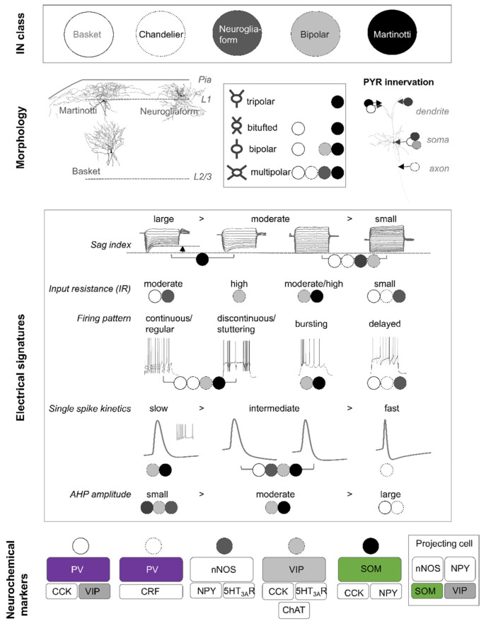
In the mammalian brain, cortical interneurons (INs) are a highly diverse group of cells. A key neurophysiological question concerns how each class of INs contributes to cortical circuit function and whether specific roles can be attributed to a selective cell type. To address this question, researchers are integrating knowledge derived from transcriptomic, histological, electrophysiological, developmental, and functional experiments to extensively characterise the different classes of INs. Our hope is that such knowledge permits the selective targeting of cell types for therapeutic endeavours. This review will focus on two of the main types of INs, namely the parvalbumin (PV+) or somatostatin (SOM+)-containing cells, and summarise the research to date on these classes.
Keywords: GABA; cortical interneurons; interneuron development; interneuron diversity; parvalbumin; somatostatin.
Conflict of interest statement
The authors declare no conflict of interest.
Figures


Similar articles
-
Diversity and Function of Somatostatin-Expressing Interneurons in the Cerebral Cortex.Int J Mol Sci. 2019 Jun 17;20(12):2952. doi: 10.3390/ijms20122952. Int J Mol Sci. 2019. PMID: 31212931 Free PMC article. Review.
-
Duration of culture and sonic hedgehog signaling differentially specify PV versus SST cortical interneuron fates from embryonic stem cells.Development. 2015 Apr 1;142(7):1267-78. doi: 10.1242/dev.111526. Development. 2015. PMID: 25804737 Free PMC article.
-
Inhibition by Somatostatin Interneurons in Olfactory Cortex.Front Neural Circuits. 2016 Aug 17;10:62. doi: 10.3389/fncir.2016.00062. eCollection 2016. Front Neural Circuits. 2016. PMID: 27582691 Free PMC article.
-
Differential effects of BMP signaling on parvalbumin and somatostatin interneuron differentiation.Development. 2009 Aug;136(15):2633-42. doi: 10.1242/dev.034439. Development. 2009. PMID: 19592576 Free PMC article.
-
Involvement of cortical fast-spiking parvalbumin-positive basket cells in epilepsy.Prog Brain Res. 2016;226:81-126. doi: 10.1016/bs.pbr.2016.04.012. Epub 2016 Jun 7. Prog Brain Res. 2016. PMID: 27323940 Free PMC article. Review.
Cited by
-
Interneuron migration impairment and brain region-specific DNA damage response following irradiation during early neurogenesis in mice.Cell Mol Life Sci. 2025 Mar 17;82(1):118. doi: 10.1007/s00018-025-05643-7. Cell Mol Life Sci. 2025. PMID: 40095026 Free PMC article.
-
Role and mechanisms of interneurons in chronic pain and pain-induced cognitive impairment.Zhong Nan Da Xue Xue Bao Yi Xue Ban. 2025 Apr 28;50(4):625-630. doi: 10.11817/j.issn.1672-7347.2025.240402. Zhong Nan Da Xue Xue Bao Yi Xue Ban. 2025. PMID: 40785676 Free PMC article. Review. Chinese, English.
-
Cell-Type-Specific Effects of Somatostatin on Synaptic Transmission in the Anterior Cingulate Cortex.J Neurosci. 2024 Mar 27;44(13):e0598232024. doi: 10.1523/JNEUROSCI.0598-23.2024. J Neurosci. 2024. PMID: 38378274 Free PMC article.
-
Loss of KCC2 in GABAergic Neurons Causes Seizures and an Imbalance of Cortical Interneurons.Front Mol Neurosci. 2022 Mar 16;15:826427. doi: 10.3389/fnmol.2022.826427. eCollection 2022. Front Mol Neurosci. 2022. PMID: 35370549 Free PMC article.
-
Translational neuronal ensembles: Neuronal microcircuits in psychology, physiology, pharmacology and pathology.Front Syst Neurosci. 2022 Aug 24;16:979680. doi: 10.3389/fnsys.2022.979680. eCollection 2022. Front Syst Neurosci. 2022. PMID: 36090187 Free PMC article. Review.
References
-
- Winnubst J., Bas E., Ferreira T.A., Wu Z., Economo M.N., Edson P., Arthur B.J., Bruns C., Rokicki K., Schauder D., et al. Reconstruction of 1000 Projection Neurons Reveals New Cell Types and Organization of Long-Range Connectivity in the Mouse Brain. Cell. 2019;179:268–281.e13. doi: 10.1016/j.cell.2019.07.042. - DOI - PMC - PubMed
-
- Riedemann T., Schmitz C., Sutor B. Immunocytochemical heterogeneity of somatostatin-expressing GABAergic interneurons in layers II and III of the mouse cingulate cortex: A combined immunofluorescence/design-based stereologic study. J. Comp. Neurol. 2015;524:2281–2299. doi: 10.1002/cne.23948. - DOI - PubMed
Publication types
MeSH terms
Substances
LinkOut - more resources
Full Text Sources
