. 2021 Aug 28;22(17):9324.
doi: 10.3390/ijms22179324.

Ptk7 Is Dynamically Localized at Neural Crest Cell-Cell Contact Sites and Functions in Contact Inhibition of Locomotion

Affiliations

Ptk7 Is Dynamically Localized at Neural Crest Cell-Cell Contact Sites and Functions in Contact Inhibition of Locomotion

Anita Grund et al. Int J Mol Sci. .

Abstract

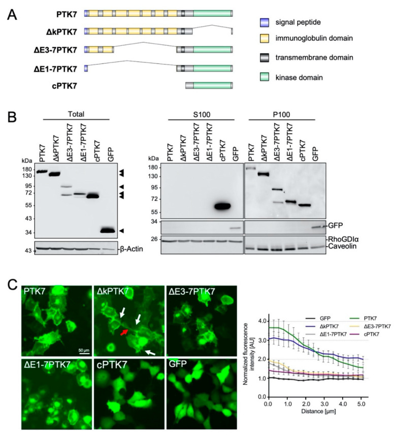
Neural crest (NC) cells are highly migratory cells that contribute to various vertebrate tissues, and whose migratory behaviors resemble cancer cell migration and invasion. Information exchange via dynamic NC cell-cell contact is one mechanism by which the directionality of migrating NC cells is controlled. One transmembrane protein that is most likely involved in this process is protein tyrosine kinase 7 (PTK7), an evolutionary conserved Wnt co-receptor that is expressed in cranial NC cells and several tumor cells. In Xenopus, Ptk7 is required for NC migration. In this study, we show that the Ptk7 protein is dynamically localized at cell-cell contact zones of migrating Xenopus NC cells and required for contact inhibition of locomotion (CIL). Using deletion constructs of Ptk7, we determined that the extracellular immunoglobulin domains of Ptk7 are important for its transient accumulation and that they mediate homophilic binding. Conversely, we found that ectopic expression of Ptk7 in non-NC cells was able to prevent NC cell invasion. However, deletion of the extracellular domains of Ptk7 abolished this effect. Thus, Ptk7 is sufficient at protecting non-NC tissue from NC cell invasion, suggesting a common role of PTK7 in contact inhibition, cell invasion, and tissue integrity.

Keywords: PTK7; cell invasion; dynamic cell–cell contacts; neural crest migration.


Conflict of interest statement

The authors declare no conflict of interest.

Figures

Figure 1
Figure 1
PTK7-GFP accumulates at cell–cell contact sites; Xenopus embryos were injected with 250 pg PTK7-GFP or 50 pg mbGFP RNA in combination with 250 pg H2B-mcherry RNA into one blastomere at the two-cell stage. NC cells were explanted at stage 17 and analyzed by spinning disk microscopy. (A) Time lapse images of PTK7-GFP expressing NC cells before (–0.8 min), during (0–7.2 min), and after (7.5 min) cell collision. The dashed square shows a higher magnification of the NC cell–cell contact zone. PTK7-GFP is highly enriched at the contact zone between NC cells, but immediately disappears when the cell–cell contacts are broken. NC cell–cell contacts are indicated by white arrows and are shown at higher magnifications in the boxed areas. Scale bar = 20 µm or 5 µm for higher magnification. (B) Time lapse images of mbGFP expressing NC cells before (–5 min), during (0–15.1 min) and after (18.5 min) collision. Like PTK7-GFP, mbGFP is localized at the membrane, however, it does not accumulate at cell–cell contact sites of colliding NC cells. (C) The graph shows the fluorescence intensity at the cell–cell contact site normalized to the membrane fluorescence at a non-contact site at 0 to 5 µm distance from the membrane. Average values and standard errors of the mean (SEM) are shown for 18 PTK7-GFP and 22 mbGFP expressing NC cells of at least three independent experiments, *** p < 0.005 (two-way ANOVA).
Figure 2
Figure 2
Localization of distinct Ptk7 deletion constructs in H1299 cells. (A) Schematic presentation of the Ptk7 domain structure and its different deletion mutants. (B) Transient expression of Ptk7-GFP variants in H1299 cells 24 h after transfection. Total cell lysates (left panel) were prepared by homogenizing the cells in RIPA-buffer and 50 µg of cell lysates were subjected to SDS-PAGE and immunoblotting. Due to the high expression level, 10 µg of lysate from cPTK7- and 5 µg from GFP-expressing cells were loaded. For subcellular localization (right panels) lysates were separated by 100,000× g centrifugation in soluble (S100) and particulate (P100) fractions. Aliquots of each fraction were analyzed by immunoblotting. GFP-containing proteins were detected by GFP-HRP antibody and ECL. Detection of β-Actin served as loading control, RhoGDIα as a marker for the cytosolic (S100) and Caveolin for the particulate (P100) fraction. (C) Fluorescence analysis of living Ptk7-GFP-expressing H1299 cells 24 h after transfection showing the subcellular localization of the transiently expressed GFP fusion proteins. Red arrows show tube-like structures, white arrows show membrane-spikes. Bar: 50 µm. The graph shows the fluorescence intensity at the cell–cell contact site normalized to the membrane fluorescence at a non-contact site at 0 to 5 µm distance from the membrane (mean ± SEM, n = 8−10).
Figure 3
Figure 3
Dynamic localization of distinct Ptk7 deletion constructs during NC cell–cell contact. Two-cell stage Xenopus embryos were injected unilaterally with 200 pg ΔkPTK7-GFP, 200 pg ΔE3-7PTK7-GFP, 200 pg ΔE1-7PTK7-GFP, or 100 pg cPTK7-GFP RNA in combination with 200 pg H2B-mcherry RNA. NC cells were explanted at stage 16–18 and protein localization was analyzed by time lapse microscopy. (A) Images show NC cells before (first column), during (second column; 0 min), and after (third column) cell–cell contact. The respective constructs are indicated for each lane. ΔkPTK7-GFP expressing NC cells showed strong protein accumulation at cell–cell contact sites and often remained connected via nanotube-like structures after they separated (red arrows). ΔE3-7PTK7-GFP, ΔE1-7PTK7-GFP, and cPTK7-GFP were not enriched at cell–cell contacts. Yellow dotted arrows indicate the direction of migrating NC cells. NC cell–cell contacts are indicated by white arrows and shown at higher magnification in the boxed areas. Scale bar = 20 µm or 5 µm for higher magnification. (B) Graph showing the average fluorescence intensity at the cell–cell contact site normalized to control sites for NC cells expressing the indicated Ptk7 deletion constructs or mbGFP. Average values and standard errors of the mean are shown for 18 PTK7-GFP, 22 mbGFP, 28 ΔkPTK7-GFP, 22 ΔE3-7PTK7-GFP, 15 ΔE1-7PTK7-GFP, and 12 cPTK7-GFP expressing NC cells of at least three independent experiments. *** p < 0.005 for ΔkPTK7-GFP versus mbGFP (two-way ANOVA). The data for mbGFP and PTK7-GFP have already been shown in Figure 1B.
Figure 4
Figure 4
∆kPTK7 overexpressing NC cells stay longer in cell–cell contact compared to controls, while single cell velocity is not affected. (A) Box plot showing cell–cell contact time (in minutes) during collision of NC cells expressing various Ptk7 constructs. NC cells expressing ΔkPTK7-GFP exhibit a significantly increased contact time compared to the control. (B) Graph blotting single cell velocity of NC cells expressing the indicated Ptk7 constructs. Box and whiskers plots summarize results of at least three independent experiments, the number of analyzed cells for each condition is indicated at the top. The box extends from the 25th to the 75th percentile, with whiskers with maximum 1.5 IQR. The median is plotted as a line inside the box. *** p < 0.005 (Kruskal–Wallis test).
Figure 5
Figure 5
Ptk7 mediates homophilic binding via its extracellular domains. (A) Schematic overview of the respective constructs. Their ability to bind to full-length Ptk7 is indicated by (+), the lack of binding by (−). HEK293 cells were transfected as indicated and immunoprecipitations were performed using anti-Myc (B) or anti-HA antibodies (C). Cell lysates are shown in the bottom panel, co-immunoprecipitated proteins in the upper panel and immunoprecipitated proteins in the middle panel. Antibodies used for Western blotting (WB) are indicated on the left, molecular weights (kDa) are indicated on the right. Representative results of at least three independent experiments are shown. The quantity of PTK7 and ∆kPTK7 co-precipitated with full-length PTK7 in respect to the respective inputs was determined by densitometric analysis (LI-COR Image Studio): mean value PTK7: 1 for ∆kPTK7: 2.6.
Figure 6
Figure 6
Loss of Ptk7 inhibits CIL in cranial NC explants. Confrontation assays were performed using explants injected with either 6.5 ng MO in combination with 100 pg mbGFP RNA or 200 pg mbRFP RNA as control. For rescue experiments 200 pg PTK7 or ∆E1-7PTK7 RNA were co-injected with the MO. (A) First column: confronted explants at the start of the experiment (time point t = 0). Second column: confronted explants at the time point of the maximal invasion (Δt). Injected constructs are indicated for each condition on the left. The overlapping area is marked by yellow fluorescence. (B) Box plot showing overlapping indexes (OI) of the different NC explants with the control. The total number of analyzed confrontation events of at least three independent experiments is indicated for each column. The box extends from the 25th to the 75th percentile, with whiskers with maximum 1.5 IQR. The median is plotted as a line inside the box. * p < 0.05, *** p < 0.005, ns.: not significant (Kruskal–Wallis test).
Figure 7
Figure 7
The extracellular domain of Ptk7 protects non-NC tissue from NC cell invasion. (AC) Embryos were injected with either 100 pg mbGFP RNA or 200 pg mbRFP RNA, alone or in combination with 500 pg PTK7, ΔkPTK7, or ΔE1-7PTK7 RNA, in one blastomere at the two-cell stage. (A) At early neurula stage, either green fluorescent NC cells or red fluorescent non-NC cells were dissected. NC cells were co-cultured with non-NC cells and analyzed by time lapse microscopy. (B) First column: confronted explants at the start of the experiment (time point t = 0). Second column: confronted explants at the time point of maximal invasion (Δt). Green fluorescent NC cells invaded red fluorescent control non-NC tissue (upper lane). Overexpression of PTK7 and ΔkPTK7 in non-NC explants prevented NC cell invasion (middle lanes). Non-NC tissue expressing ΔE1-7PTK7 was invaded by NC cells (bottom panel) as it was observed in the control. Scale bar = 100 µm. (C) Box plot showing overlapping indexes (OI) of NC explants confronted with non-NC tissue expressing mbRFP alone or in combination with various Ptk7 deletion constructs. Overexpression of PTK7 and ΔkPTK7, but not ΔE1-7PTK7, significantly decreased the OI. Number of analyzed confrontation events of at least three independent experiments is indicated for each column. The box extends from the 25th to the 75th percentile, with whiskers with maximum 1.5 IQR. The median is plotted as a line inside the box. * p < 0.05, *** p < 0.005 (Kruskal–Wallis test). (D) Marker expression of the explanted tissue was determined by performing RT-PCR utilizing the NC marker sox10, ap2α, and twist as well as the endoderm marker sox17α. Amplification of histone H4 (H4) was used as an internal control.

References

    1. Szabo A., Mayor R. Mechanisms of Neural Crest Migration. Annu. Rev. Genet. 2018;52:43–63. doi: 10.1146/annurev-genet-120417-031559. - DOI - PubMed
    1. Shellard A., Mayor R. Integrating chemical and mechanical signals in neural crest cell migration. Curr. Opin. Genet. Dev. 2019;57:16–24. doi: 10.1016/j.gde.2019.06.004. - DOI - PMC - PubMed
    1. Theveneau E., Mayor R. Neural crest migration: Interplay between chemorepellents, chemoattractants, contact inhibition, epithelial-mesenchymal transition, and collective cell migration. Wiley Interdiscip. Rev. Dev. Biol. 2012;1:435–445. doi: 10.1002/wdev.28. - DOI - PubMed
    1. Kuriyama S., Mayor R. Molecular analysis of neural crest migration. Philos Trans. R Soc. Lond. B Biol. Sci. 2008;363:1349–1362. doi: 10.1098/rstb.2007.2252. - DOI - PMC - PubMed
    1. Gallik K.L., Treffy R.W., Nacke L.M., Ahsan K., Rocha M., Green-Saxena A., Saxena A. Neural crest and cancer: Divergent travelers on similar paths. Mech. Dev. 2017;148:89–99. doi: 10.1016/j.mod.2017.08.002. - DOI - PMC - PubMed

Substances