Review
. 2021 Sep 1;22(17):9502.
doi: 10.3390/ijms22179502.

RNA Granules in the Mitochondria and Their Organization under Mitochondrial Stresses

Affiliations
Review

RNA Granules in the Mitochondria and Their Organization under Mitochondrial Stresses

Vanessa Joanne Xavier et al. Int J Mol Sci. .

Abstract

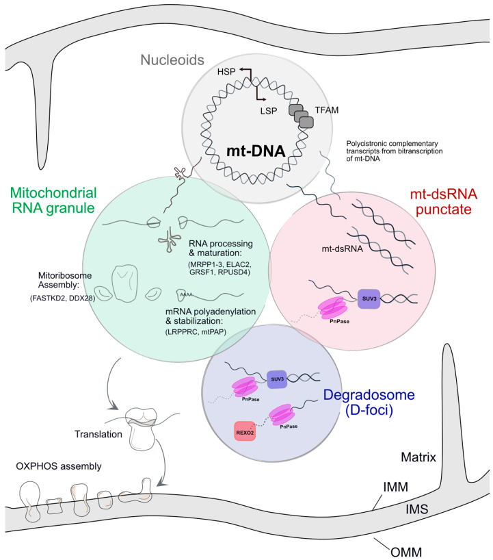
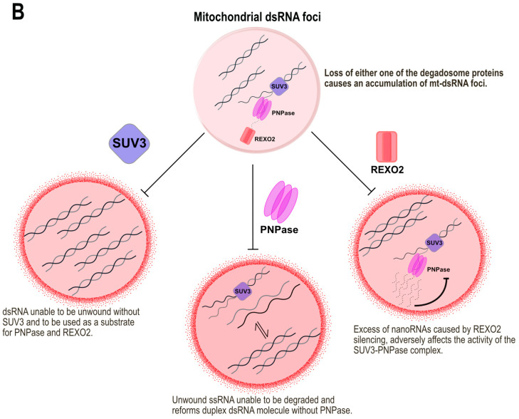
The human mitochondrial genome (mtDNA) regulates its transcription products in specialised and distinct ways as compared to nuclear transcription. Thanks to its mtDNA mitochondria possess their own set of tRNAs, rRNAs and mRNAs that encode a subset of the protein subunits of the electron transport chain complexes. The RNA regulation within mitochondria is organised within specialised, membraneless, compartments of RNA-protein complexes, called the Mitochondrial RNA Granules (MRGs). MRGs were first identified to contain nascent mRNA, complexed with many proteins involved in RNA processing and maturation and ribosome assembly. Most recently, double-stranded RNA (dsRNA) species, a hybrid of the two complementary mRNA strands, were found to form granules in the matrix of mitochondria. These RNA granules are therefore components of the mitochondrial post-transcriptional pathway and as such play an essential role in mitochondrial gene expression. Mitochondrial dysfunctions in the form of, for example, RNA processing or RNA quality control defects, or inhibition of mitochondrial fission, can cause the loss or the aberrant accumulation of these RNA granules. These findings underline the important link between mitochondrial maintenance and the efficient expression of its genome.

Keywords: RNA degradation; RNA processing; degradosome; dsRNA; liquid–liquid phase separation (LLPS); mitochondrial RNA granules (MRGs); mitochondrial gene expression; nucleoids.


Conflict of interest statement

The authors declare no conflict of interest.

Figures

Figure 1
Figure 1
Nucleoids containing a single mt-DNA molecule, condensed by TFAM, transcribe polycistronic mRNAs. A single-stranded mRNA transcript is thought to undergo endonucleolytic processing and maturation in the Mitochondrial RNA granule (MRG). It is also the site of mitoribosome assembly. Matured mRNA transcripts transit out of the MRGs to be translated into subunits of the ETC. Bidirectional transcription of the mt-DNA from its Heavy strand promoter (HSP) and Light strand promoter (LSP) can lead to the complementary transcripts forming duplex mt-dsRNA molecules which have been observed in foci similar to MRGs. mt-dsRNA is mainly regulated by the degradosome proteins, SUV3-PNPase. The Degradosome (D-foci) is the site of mt-RNA degradation by SUV3, PNPase and REXO2. While colocalization between Nucleoids, MRGs and D-foci has been demonstrated, the association of mt-dsRNA granules to these structures has yet to be further investigated. Either they are separate entities or part of the MRGs, which would imply that the different structures are tightly connected. IMM: Inner mitochondrial membrane, IMS: Intermembrane space, OMM: Outer mitochondrial membrane.
Figure 2
Figure 2
Consequences on the structure of MRGs and mt-dsRNA foci when its components are dysregulated. (A) Various bona fide MRG proteins phase out into liquid droplets, surrounding an RNA core in MRGs. The silencing of any one of these proteins does not destroy the persistence of MRGs in the matrix. The loss of the crucial nascent RNA core leads to the dissolution of MRGs. However, in this case, the MRG proteins can still be found diffused throughout the matrix when visualized by immunofluorescence. Enlarged mitobulbs of mitochondria are the result of disrupted membrane dynamics. Nucleoids and MRGs form tight clusters within them while maintaining their individual structures. (B) The loss of either one of the degradosome proteins of SUV3, PNPase and REXO2 causes an accumulation of mt-dsRNA foci in the matrix. The silencing of each of these proteins results in a different mechanism leading to the accumulation of mt-dsRNA relating to its function.
Figure 2
Figure 2
Consequences on the structure of MRGs and mt-dsRNA foci when its components are dysregulated. (A) Various bona fide MRG proteins phase out into liquid droplets, surrounding an RNA core in MRGs. The silencing of any one of these proteins does not destroy the persistence of MRGs in the matrix. The loss of the crucial nascent RNA core leads to the dissolution of MRGs. However, in this case, the MRG proteins can still be found diffused throughout the matrix when visualized by immunofluorescence. Enlarged mitobulbs of mitochondria are the result of disrupted membrane dynamics. Nucleoids and MRGs form tight clusters within them while maintaining their individual structures. (B) The loss of either one of the degradosome proteins of SUV3, PNPase and REXO2 causes an accumulation of mt-dsRNA foci in the matrix. The silencing of each of these proteins results in a different mechanism leading to the accumulation of mt-dsRNA relating to its function.

References

    1. Zhao R.Z., Jiang S., Zhang L., Yu Z. Bin Mitochondrial electron transport chain, ROS generation and uncoupling (Review) Int. J. Mol. Med. 2019;44:3–15. doi: 10.3892/ijmm.2019.4188. - DOI - PMC - PubMed
    1. Anderson S., Bankier A.T., Barrell B.G., de Bruijn M.H.L., Coulson A.R., Drouin J., Eperon I.C., Nierlich D.P., Roe B.A., Sanger F. Sequence and organization of the human mitochondrial genome. Nature. 1981;290:1–18. doi: 10.1038/290457a0. - DOI - PubMed
    1. Aloni Y., Attardi G. Symmetrical in vivo transcription of mitochondrial DNA in HeLa cells. Proc. Natl. Acad. Sci. USA. 1971;68:1757–1761. doi: 10.1073/pnas.68.8.1757. - DOI - PMC - PubMed
    1. Aloni Y., Attardi G. Expression of the Mitochondrial Genome in Hela Cells. J. Cell Biol. 1971;56:819–831. doi: 10.1083/jcb.56.3.819. - DOI - PMC - PubMed
    1. Gustafsson C.M., Falkenberg M., Larsson N.G. Maintenance and Expression of Mammalian Mitochondrial DNA. Annu. Rev. Biochem. 2016;85:133–160. doi: 10.1146/annurev-biochem-060815-014402. - DOI - PubMed

MeSH terms

LinkOut - more resources