

The Role of the α Cell in the Pathogenesis of Diabetes: A World beyond the Mirror
- PMID: 34502413
- PMCID: PMC8431704
- DOI: 10.3390/ijms22179504
The Role of the α Cell in the Pathogenesis of Diabetes: A World beyond the Mirror
Abstract
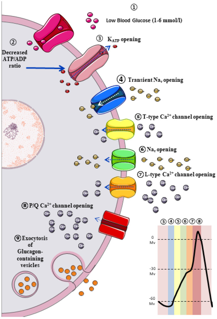
Type 2 Diabetes Mellitus (T2DM) is one of the most prevalent chronic metabolic disorders, and insulin has been placed at the epicentre of its pathophysiological basis. However, the involvement of impaired alpha (α) cell function has been recognized as playing an essential role in several diseases, since hyperglucagonemia has been evidenced in both Type 1 and T2DM. This phenomenon has been attributed to intra-islet defects, like modifications in pancreatic α cell mass or dysfunction in glucagon's secretion. Emerging evidence has shown that chronic hyperglycaemia provokes changes in the Langerhans' islets cytoarchitecture, including α cell hyperplasia, pancreatic beta (β) cell dedifferentiation into glucagon-positive producing cells, and loss of paracrine and endocrine regulation due to β cell mass loss. Other abnormalities like α cell insulin resistance, sensor machinery dysfunction, or paradoxical ATP-sensitive potassium channels (KATP) opening have also been linked to glucagon hypersecretion. Recent clinical trials in phases 1 or 2 have shown new molecules with glucagon-antagonist properties with considerable effectiveness and acceptable safety profiles. Glucagon-like peptide-1 (GLP-1) agonists and Dipeptidyl Peptidase-4 inhibitors (DPP-4 inhibitors) have been shown to decrease glucagon secretion in T2DM, and their possible therapeutic role in T1DM means they are attractive as an insulin-adjuvant therapy.
Keywords: Langerhans’ islets; glucagon; hyperglycaemia; hypoglycaemia; type 2 diabetes.
Conflict of interest statement
The authors declare no conflict of interest.
Figures
Similar articles
-
Activation of Transmembrane Bile Acid Receptor TGR5 Modulates Pancreatic Islet α Cells to Promote Glucose Homeostasis.J Biol Chem. 2016 Mar 25;291(13):6626-40. doi: 10.1074/jbc.M115.699504. Epub 2016 Jan 12. J Biol Chem. 2016. PMID: 26757816 Free PMC article.
-
Long-term liraglutide treatment is associated with increased insulin content and secretion in β-cells, and a loss of α-cells in ZDF rats.Pharmacol Res. 2013 Oct;76:58-66. doi: 10.1016/j.phrs.2013.07.005. Epub 2013 Jul 26. Pharmacol Res. 2013. PMID: 23891763
-
Human islets contain a subpopulation of glucagon-like peptide-1 secreting α cells that is increased in type 2 diabetes.Mol Metab. 2020 Sep;39:101014. doi: 10.1016/j.molmet.2020.101014. Epub 2020 May 12. Mol Metab. 2020. PMID: 32413586 Free PMC article.
-
Improved glucose regulation in type 2 diabetic patients with DPP-4 inhibitors: focus on alpha and beta cell function and lipid metabolism.Diabetologia. 2016 May;59(5):907-17. doi: 10.1007/s00125-016-3899-2. Epub 2016 Feb 19. Diabetologia. 2016. PMID: 26894277 Review.
-
Preproglucagon Products and Their Respective Roles Regulating Insulin Secretion.Endocrinology. 2021 Oct 1;162(10):bqab150. doi: 10.1210/endocr/bqab150. Endocrinology. 2021. PMID: 34318874 Free PMC article. Review.
Cited by
-
Real-World Comparative Evaluation of Add-On Glucagon-like Peptide 1 Receptor Agonist in Type 2 Diabetes Treated with or without Insulin.Pharmaceuticals (Basel). 2022 Dec 15;15(12):1569. doi: 10.3390/ph15121569. Pharmaceuticals (Basel). 2022. PMID: 36559020 Free PMC article.
-
An Update on the Current and Emerging Use of Thiazolidinediones for Type 2 Diabetes.Medicina (Kaunas). 2022 Oct 17;58(10):1475. doi: 10.3390/medicina58101475. Medicina (Kaunas). 2022. PMID: 36295635 Free PMC article. Review.
-
A Supportive Role of Mesenchymal Stem Cells on Insulin-Producing Langerhans Islets with a Specific Emphasis on The Secretome.Biomedicines. 2023 Sep 18;11(9):2558. doi: 10.3390/biomedicines11092558. Biomedicines. 2023. PMID: 37761001 Free PMC article. Review.
-
Induction of diabetes by Tacrolimus in a phenotypic model of obesity and metabolic syndrome.Front Endocrinol (Lausanne). 2024 Apr 29;15:1388361. doi: 10.3389/fendo.2024.1388361. eCollection 2024. Front Endocrinol (Lausanne). 2024. PMID: 38745946 Free PMC article.
-
Pancreatic β-cell senescence in diabetes: mechanisms, markers and therapies.Front Endocrinol (Lausanne). 2023 Aug 31;14:1212716. doi: 10.3389/fendo.2023.1212716. eCollection 2023. Front Endocrinol (Lausanne). 2023. PMID: 37720527 Free PMC article. Review.
References
-
- Noncommunicable Diseases Progress Monitor 2017. [(accessed on 21 March 2021)]; Available online: https://www.who.int/publications-detail-redirect/9789241513029.
-
- IDF Diabetes Atlas 9th Edition 2019. [(accessed on 21 March 2021)]; Available online: https://www.diabetesatlas.org/en/
Publication types
MeSH terms
Substances
LinkOut - more resources
Full Text Sources
Medical
Miscellaneous
