. 2021 Sep 1;22(17):9514.
doi: 10.3390/ijms22179514.

Docking to a Basic Helix Promotes Specific Phosphorylation by G1-Cdk1

Affiliations

Docking to a Basic Helix Promotes Specific Phosphorylation by G1-Cdk1

Ilona Faustova et al. Int J Mol Sci. .

Abstract

Cyclins are the activators of cyclin-dependent kinase (CDK) complex, but they also act as docking scaffolds for different short linear motifs (SLiMs) in CDK substrates and inhibitors. According to the unified model of CDK function, the cell cycle is coordinated by CDK both via general CDK activity thresholds and cyclin-specific substrate docking. Recently, it was found that the G1-cyclins of S. cerevisiae have a specific function in promoting polarization and growth of the buds, making the G1 cyclins essential for cell survival. Thus, while a uniform CDK specificity of a single cyclin can be sufficient to drive the cell cycle in some cells, such as in fission yeast, cyclin specificity can be essential in other organisms. However, the known G1-CDK specific LP docking motif, was not responsible for this essential function, indicating that G1-CDKs use yet other unknown docking mechanisms. Here we report a discovery of a G1 cyclin-specific (Cln1,2) lysine-arginine-rich helical docking motif (the K/R motif) in G1-CDK targets involved in the mating pathway (Ste7), transcription (Xbp1), bud morphogenesis (Bud2) and spindle pole body (Spc29, Spc42, Spc110, Sli15) function of S. cerevisiae. We also show that the docking efficiency of K/R motif can be regulated by basophilic kinases such as protein kinase A. Our results further widen the list of cyclin specificity mechanisms and may explain the recently demonstrated unique essential function of G1 cyclins in budding yeast.

Keywords: SLiM; cyclin specificity; cyclin-dependent kinase; kinase specificity; phosphorylation.


Conflict of interest statement

The authors declare no conflict of interest.

Figures

Figure 1
Figure 1
A substrate docking interaction different from the known LP interaction, governs the phosphorylation specificity of a group of key targets of G1-Cdk1. 32P-autoradiographs showing phosphorylation of Cdk1 targets by the four major cyclin-Cdk1 complexes and the LP docking deficient Cln2(lpd)-Cdk1. +2K/R:+3K/R shows the number Cdk1 consensus phosphorylation sites with basic residue in +2 position and +3 position. The experiments were performed twice, a representative example is shown.
Figure 2
Figure 2
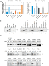
Clusters of basic residues promote phosphorylation by G1-Cdk1. (A) 32P-autoradiographs of phosphorylation reactions of Sli15 fragment 421–511 with all phosphorylation sites (upper panel) or with just S448 as the only Cdk1 consensus site by different cyclin-Cdk1 complexes in vitro. (B) Sequence of Sli15 positions 448–511 showing the truncations and mutations used in panel C. (C) Phospho-images showing phosphorylation of the Sli15 mutants by Cln2-Cdk1. The mutants are described in ‘B’. In the kr mutant, all highlighted K/R residues are mutated to N/Q residues, respectively. (D) Sequences of the predicted α-helical K/R docking motifs in Bud2 and Rtt109. (E) 32P-autoradiographs showing the phosphorylation of Bud2 and Rtt109 proteins by Cln2- and Clb2-Cdk1. wt/h shows the relative phosphorylation rate of the wild-type substrate protein compared to the K/R docking site mutant. (F) Helical wheel representations of the K/R docking motifs in Sli15, Bud2 and Rtt109 show the clustering of basic residues on one side of the α-helix. (G) Multiple sequence alignment of K/R motifs in Sli15, Bud2 and Rtt109.
Figure 3
Figure 3
Cln2 mutants deficient in either LP or K/R docking. (A) The docking specificity of mutant Cln2-Cdk1 complexes was analyzed in vitro by measuring phosphorylation of LP-dependent substrate Sic1-S76 and K/R-dependent Spc42. The mutations in each Cln2 mutant and their position on the Cln2 surface are shown in Figure S2A,B. Cln2-ΔC indicates deletion of the C-terminal intrinsically disordered domain of Cln2 (positions 372–545). (B) 32P-autoradiographs showing the phosphorylation of histone H1, Sic1ΔC, Whi5, Sli15(421–511 S448) and Sli15(421–511 S448; kr), where the basic residues involved in K/R docking (highlighted in Figure 2B) are mutated to Q and N, by different Cln2-Cdk1 mutant complexes. (C) The effect of K/R docking on phosphorylation of various G1-Cdk1 targets was studied in vitro using Cln2(krd2) mutant. Representative 32P-autoradiographs are shown.
Figure 4
Figure 4
K/R docking mediates Cln2 function in bud morphogenesis and spindle pole body dynamics. (A) The time from Start, measured by the nuclear export of Whi5-mCherry, to budding was studied in time-lapse microscopy in single cells of cln1Δ strain expressing the indicated CLN2 variant from CLN2 promoter. The error bars show 95% confidence intervals of the median. * indicates p-value < 0.05 and ** p-value < 0.01 in pairwise comparisons of the indicated condition with CLN2(wt) by Mann-Whitney U-test. (B) Microscopy images showing the effect of GST-Cln2 overexpression on bud morphology. (C) Plot showing the capability of different Cln2 variants to promote elongated bud growth. The bud length was measured 2 h after PGAL1-CLN2 induction. **** indicates p-value < 0.0001 for pairwise comparisons with CLN2(wt) using Mann-Whitney U-test. (D) Multisite phosphorylation of Cdc24-13myc was studied by Phos-tag Western blotting of cln1Δ strains expressing the indicated CLN2 variant from CLN2 promoter. The cultures were synchronized in G1 using α-factor and were released to the cell cycle. Cdc24 phosphorylation was analyzed 30 and 45 min after releasing from α-factor arrest, at the peak of Cln2 expression. (E) The fraction of cells with short or long spindles was studied with fluorescence microscopy in cln1Δ strains expressing different Cln2 mutants, Spc42-EGFP and Tub1-mCherry. ** denotes p < 0.01 for comparison with CLN2wt by χ2-test.
Figure 5
Figure 5
K/R docking regulates Sli15 localization dynamics. (A) Images showing the subcellular localization of Sli15-GFP during the cell cycle in cells expressing Cln2(wt) or Cln2(krd2). The nuclear localization of Whi5-mCherry was used as the reference for cell cycle progression. (B) Plots showing the fraction of cells with the indicated Sli15-GFP localization. Early G1 cells were unbudded cells with nuclear Whi5-mCherry, whereas late G1 cells were unbudded but without nuclear Whi5-mCherry signal. Small budded cells were denoted as S phase and large budded cells were categorized as G2/M cells. The experiments were performed using cln1Δ cln2Δ strains expressing the indicated CLN2 variant under CLN2 promoter on a centromeric plasmid.
Figure 6
Figure 6
Phosphorylation adjacent to the K/R docking α-helix switches the docking off. (A) Scheme showing the sequence of Bud2 C terminus with two potential protein kinase A (PKA) phosphorylation sites in the K/R docking motif. (B) 32P-autoradiographs showing the phosphorylation of T1092 and T1101 by PKA. (C) The C terminus of Bud2 was first phosphorylated by PKA without 32P-ATP under conditions where Bud2 is expected to be fully phosphorylated at T1092 and T1101 by PKA, followed by removal of PKA and subsequent phosphorylation by Cln2-Cdk1 in the presence of 32P-ATP. “−“ indicates no pre-phosphorylation of Bud2ΔN, whereas “+” indicates that Bud2 ΔN has been pre-phosphorylated by PKA. An autoradiograph showing the inhibition of Bud2 phosphorylation by Cln2-Cdk1 by pre-phosphorylation of the substrate with PKA. (D) The effect of Bud2 pre-phosphorylation by PKA on subsequent phosphorylation by Cln2- and Clb2-Cdk1. The error bars show standard deviation.

Similar articles

Cited by

References

    1. Morgan D.O. The Cell Cycle: Principles of Control. New Science Press; London, UK: 2007.
    1. Enserink J.M., Kolodner R.D. An overview of Cdk1-controlled targets and processes. Cell Div. 2010;5:11. doi: 10.1186/1747-1028-5-11. - DOI - PMC - PubMed
    1. Stern B., Nurse P. A quantitative model for the cdc2 control of S phase and mitosis in fission yeast. Trends Genet. 1996;12:345–350. doi: 10.1016/S0168-9525(96)80016-3. - DOI - PubMed
    1. Swaffer M.P., Jones A.W., Flynn H.R., Snijders A.P., Nurse P. CDK Substrate Phosphorylation and Ordering the Cell Cycle. Cell. 2016;167:1750–1761.e16. doi: 10.1016/j.cell.2016.11.034. - DOI - PMC - PubMed
    1. Fisher D.L., Nurse P. A single fission yeast mitotic cyclin B p34cdc2 kinase promotes both S-phase and mitosis in the absence of G1 cyclins. EMBO J. 1996;15:850–860. doi: 10.1002/j.1460-2075.1996.tb00420.x. - DOI - PMC - PubMed

MeSH terms

Substances