. 2021 Sep 2;22(17):9536.
doi: 10.3390/ijms22179536.

Novel Lytic Enzyme of Prophage Origin from Clostridium botulinum E3 Strain Alaska E43 with Bactericidal Activity against Clostridial Cells

Affiliations

Novel Lytic Enzyme of Prophage Origin from Clostridium botulinum E3 Strain Alaska E43 with Bactericidal Activity against Clostridial Cells

Agnieszka Morzywolek et al. Int J Mol Sci. .

Abstract

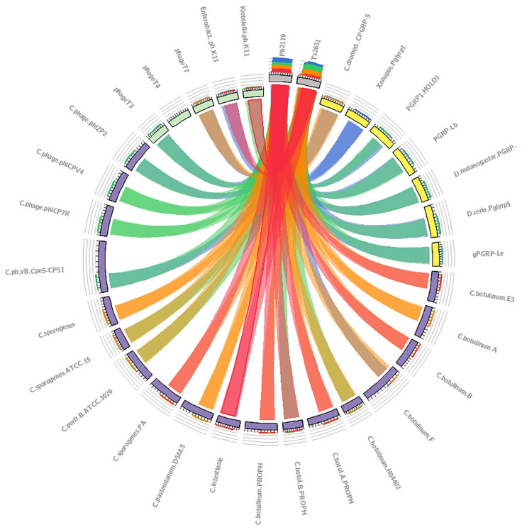
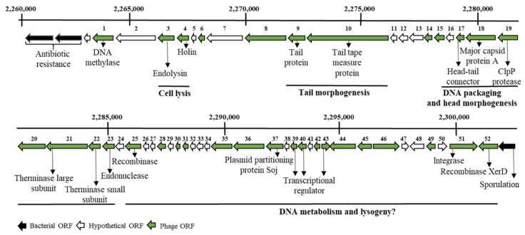
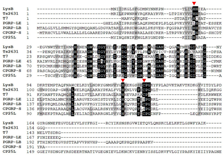
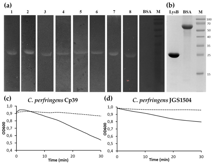
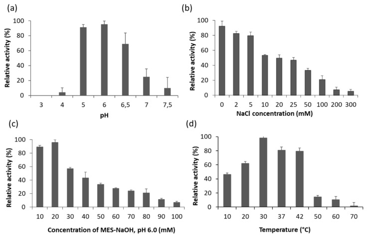
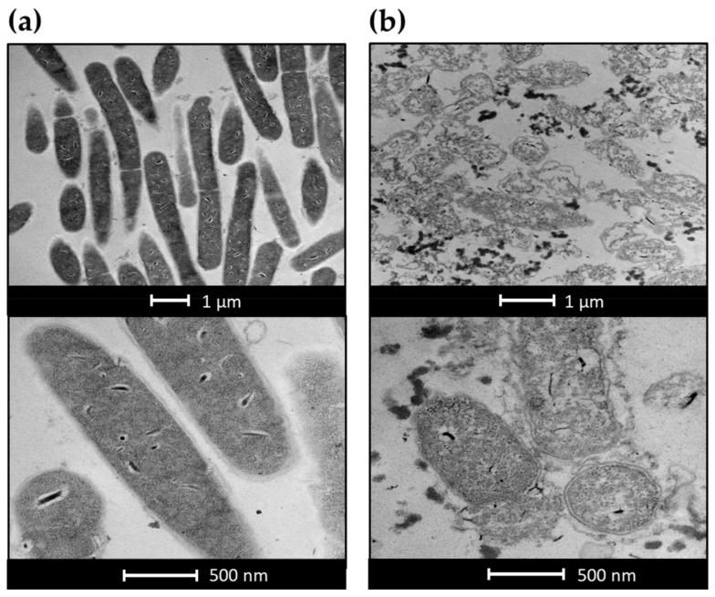
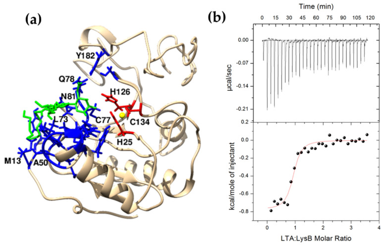
Clostridium botulinum is a Gram-positive, anaerobic, spore-forming bacterium capable of producing botulinum toxin and responsible for botulism of humans and animals. Phage-encoded enzymes called endolysins, which can lyse bacteria when exposed externally, have potential as agents to combat bacteria of the genus Clostridium. Bioinformatics analysis revealed in the genomes of several Clostridium species genes encoding putative N-acetylmuramoyl-l-alanine amidases with anti-clostridial potential. One such enzyme, designated as LysB (224-aa), from the prophage of C. botulinum E3 strain Alaska E43 was chosen for further analysis. The recombinant 27,726 Da protein was expressed and purified from E. coli Tuner(DE3) with a yield of 37.5 mg per 1 L of cell culture. Size-exclusion chromatography and analytical ultracentrifugation experiments showed that the protein is dimeric in solution. Bioinformatics analysis and results of site-directed mutagenesis studies imply that five residues, namely H25, Y54, H126, S132, and C134, form the catalytic center of the enzyme. Twelve other residues, namely M13, H43, N47, G48, W49, A50, L73, A75, H76, Q78, N81, and Y182, were predicted to be involved in anchoring the protein to the lipoteichoic acid, a significant component of the Gram-positive bacterial cell wall. The LysB enzyme demonstrated lytic activity against bacteria belonging to the genera Clostridium, Bacillus, Staphylococcus, and Deinococcus, but did not lyse Gram-negative bacteria. Optimal lytic activity of LysB occurred between pH 4.0 and 7.5 in the absence of NaCl. This work presents the first characterization of an endolysin derived from a C. botulinum Group II prophage, which can potentially be used to control this important pathogen.

Keywords: Clostridium botulinum; N-acetylmuramoyl-l-alanine amidase; endolysin; lipoteichoic acid; prophage.


Conflict of interest statement

The authors declare no conflict of interest.

Figures

Figure 1
Figure 1
The similarity analysis of thermostable endolysins Ph2119 and Ts2631 compared to phage and bacterial lytic enzymes including Clostridium enzymes and eukaryotic proteins recognizing peptidoglycan (PGRPs), visualized using Circoletto software [33]. The Basic Local Alignment Search Tool (BLAST) sequence comparison results are represented in four quartiles, each shown in a different color pattern. Red ribbons reflect the highest score, corresponding to 33–34% amino acid sequence identity, orange and green indicate medium scores, and blue indicates the lowest percentage of identity (26%). The width of the ribbons represents alignment length. Original dataset with the protein amino acid sequences and their respective GenBank or Protein Data Bank (PDB) accessions numbers and the BLAST results with E values are available as Supplementary Materials S1 and S2.
Figure 2
Figure 2
A fragment of C. botulinum E3 strain Alaska E43 genome map with predicted open reading frames (ORFs) within the prophage region. ORFs are depicted as arrows in the expected direction of transcription. Gray: bacterial ORF, green: phage ORF, white: hypothetical ORF. Putative functions were assigned based on conserved domain searches. The scheme was performed based on a graphical view of the complete genome sequence of C. botulinum E3 strain Alaska E43 (https://www.ncbi.nlm.nih.gov/nuccore/CP001078.1?report=graph; Accessed on 25 May 2021).
Figure 3
Figure 3
Multiple alignment of the amino acid sequence of LysB endolysin shows its homology to Ts2631 endolysin, T7 lysozyme, and eukaryotic peptidoglycan recognition proteins (PGRPs). GenBank or PDB accession numbers are LysB (ACD52487), Ts2631 from T. scotoductus phage vB_Tsc2631 (AIM47292.1), T7 from enterobacteria phage T7 (AAB32819.1), gPGRP-LE from D. melanogaster (PDB entry: 2CB3_B), PGRP-LB from D. melanogaster (PDB entry: 1OHT_A), CPGRP-S from C. dromedarius (PDB entry: 3O4K), CP25L from C. perfringens phage vB_CpeS-CP51 (AGH27916.1). The red triangles indicate residues responsible for Zn2+ binding (H25, H126, C134). The alignment was generated with the Clustal Omega program with default options. The black background represents 100% amino acid sequence identity, while the gray background indicates amino acid conservation at 70%.
Figure 4
Figure 4
Overproduction and purification of LysB endolysin. (a) Overproduction in E. coli Tuner(DE3) cells was carried out with 1 mM IPTG for 4 h at both 37 °C and 30 °C or overnight at 18 °C. Then cells were harvested, and after sonication, cell lysates were centrifuged to obtain clear supernatant with soluble proteins (S) and pellet (P) fractions. (b) Purified LysB endolysin. All samples were mixed with Laemmli buffer and loaded on 12.5% SDS-PAGE. Gels were stained with Coomassie Brilliant Blue R-250. (M) Protein standards where values at left indicate molecular masses (in kDa) (Thermo Fisher Scientific, Waltham, MA USA).
Figure 5
Figure 5
Lytic activity of LysB endolysin. (a) Zymogram containing (1) C. sporogenes ATCC 7955, (2) C. intestinale ATCC 49213, (3) B. cereus ATCC 13061, (4) B. megaterium ATCC 14581, (5) B. mycoides KPD 15, (6) B. thuringensis KPD 114, (7) S. aureus ATCC 25923, (8) D. radiodurans ATCC 13939, and bovine serum albumin (BSA). (b) SDS-PAGE (12.5%) analysis of purified LysB endolysin (LysB) and BSA as a quantity control; results of turbidity reduction assay against (c) C. perfringens Cp39 and (d) C. perfringens JGS1504. The dotted lines indicate controls (cells without endolysin), and solid lines indicate bacterial cells incubated at 22 °C with LysB at a concentration of 100 µg/mL. The data shown represent one of three independent experiments. (M) Protein standards where values at right indicate molecular masses (in kDa) (Thermo Fisher Scientific, Waltham, MA, USA).
Figure 6
Figure 6
Effects of (a) pH; (b) NaCl; (c) the optimal value of the buffer concentration; and (d) temperature on the lytic activity of LysB against C. sporogenes ATCC 7955 cells. Relative activity was calculated by comparing the lytic activity at a specific condition with the maximal lytic activity within the dataset. Each experiment was repeated in triplicate, error bars indicate the standard deviations.
Figure 7
Figure 7
Transmission electron microscopy of LysB-treated C. sporogenes ATCC 7955. (a) Untreated C. sporogenes cells, (b) C. sporogenes treated with 50 µg/mL LysB for 20 min.
Figure 8
Figure 8
Size-exclusion chromatography and sedimentation velocity analytical ultracentrifugation analysis of LysB. (a) Elution profile of LysB endolysin on Superdex 75 sizing column. LysB endolysin (3 mg/mL) in a final volume of 300 µL was loaded on Superdex 75 10/300 GL (AKTA Pure chromatography system, GE Healthcare, Little Chalfont, UK) and eluted at a flow rate of 0.8 mL/min in buffer consisting of 25 mM NaH2PO4, 150 mM NaCl, pH 8.0. Elution of LysB endolysin was detected by absorption at 280 nm (mAU, milli-absorbance units). The position of molecular mass standards are shown as arrows: dextran blue: 2000 kDa, bovine serum albumin: 66 kDa, trypsin inhibitor: 20 kDa, cytochrome C: 12.4 kDa, aprotinin: 6.5 kDa. (b) Sedimentation velocity data were overlaid with the best-fit curves (lines) obtained from sedimentation coefficient distribution analysis. For the clarity of the drawing, only every fifth scan and fifth data point are shown. Below, residuals of the experimental fits. (c) Sedimentation coefficient distributions (c(s)) of LysB endolysin. Peak 1 corresponds to LysB monomer; peak 2 corresponds to LysB homodimer.
Figure 9
Figure 9
Interaction between LysB and lipoteichoic acid. (a) Structural model of LysB endolysin (template: PDB 3O4K). The residues M13, H43, N47, G48 W49, A50, L73, A75, H76, Q78, N81, and Y182 form the LTA binding site (3.5 Å blue zone from green LTA). H25, H126, and C 134 (red) bind Zn2+ ion (yellow). For the clarity of the presentation, only one molecule from the LysB homodimer is shown. (b) Calorimetric titration isotherm of the binding interaction between LTA and LysB in 20 mM potassium phosphate buffer, pH 8.0, 10% glycerol at 25 °C.

Similar articles

Cited by

References

    1. Shukla H.D., Sharma S.K. Clostridium botulinum: A bug with beauty and weapon. Crit. Rev. Microbiol. 2005;31:11–18. doi: 10.1080/10408410590912952. - DOI - PubMed
    1. Poulain B., Popoff M.R. Why Are Botulinum Neurotoxin-Producing Bacteria So Diverse and Botulinum Neurotoxins So Toxic? Toxins. 2019;11:34. doi: 10.3390/toxins11010034. - DOI - PMC - PubMed
    1. Sobel J. Botulism. Clin. Infect. Dis. 2005;41:1167–1173. doi: 10.1086/444507. - DOI - PubMed
    1. Mazuet C., Legeay C., Sautereau J., Ma L., Bouchier C., Bouvet P., Popoff M.R. Diversity of Group I and II Clostridium botulinum Strains from France Including Recently Identified Subtypes. Genome Biol. Evol. 2016;8:1643–1660. doi: 10.1093/gbe/evw101. - DOI - PMC - PubMed
    1. Lonati D., Schicchi A., Crevani M., Buscaglia E., Scaravaggi G., Maida F., Cirronis M., Petrolini V.M., Locatelli C.A. Foodborne Botulism: Clinical Diagnosis and Medical Treatment. Toxins. 2020;12:509. doi: 10.3390/toxins12080509. - DOI - PMC - PubMed

MeSH terms

Supplementary concepts

LinkOut - more resources