Review
. 2021 Sep 1;21(17):5891.
doi: 10.3390/s21175891.

Multiphoton Laser Fabrication of Hybrid Photo-Activable Biomaterials

Affiliations
Review

Multiphoton Laser Fabrication of Hybrid Photo-Activable Biomaterials

Margaux Bouzin et al. Sensors (Basel). .

Abstract

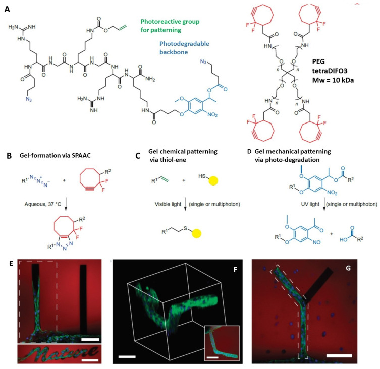
The possibility to shape stimulus-responsive optical polymers, especially hydrogels, by means of laser 3D printing and ablation is fostering a new concept of "smart" micro-devices that can be used for imaging, thermal stimulation, energy transducing and sensing. The composition of these polymeric blends is an essential parameter to tune their properties as actuators and/or sensing platforms and to determine the elasto-mechanical characteristics of the printed hydrogel. In light of the increasing demand for micro-devices for nanomedicine and personalized medicine, interest is growing in the combination of composite and hybrid photo-responsive materials and digital micro-/nano-manufacturing. Existing works have exploited multiphoton laser photo-polymerization to obtain fine 3D microstructures in hydrogels in an additive manufacturing approach or exploited laser ablation of preformed hydrogels to carve 3D cavities. Less often, the two approaches have been combined and active nanomaterials have been embedded in the microstructures. The aim of this review is to give a short overview of the most recent and prominent results in the field of multiphoton laser direct writing of biocompatible hydrogels that embed active nanomaterials not interfering with the writing process and endowing the biocompatible microstructures with physically or chemically activable features such as photothermal activity, chemical swelling and chemical sensing.

Keywords: 3D printing; hydrogels; photo-ablation; photo-polymerization.


Conflict of interest statement

The authors declare no conflict of interest.

Figures

Figure 1
Figure 1
Norrish type of photoinitiation of the radical polymerization for type I photoinitiators (A) and type II photoinitiators (B). (C) represents schematically the photoinitiation of the PI LAP (see Table 1). (D) represents schematically the photoinitiation of one common photo-click reaction based on the norbornene moiety. (E) represents one common photo-cleavage reaction involving the NBE group (see Table 1).
Figure 2
Figure 2
Schematics of the general mechanisms at the origin of the energy deposition in polymeric materials. Excitation into the conduction band (ionization) leads to quasi-free electrons still loosely bound to atoms. The excitation energy into the conduction band can be provided either by MPI or TI, depending on the Keldysh parameter, or by impact ionization from an energetic quasi-free electron that has gained its energy through photon absorption in a non-resonant process called ‘inverse Bremsstrahlung’ in the course of collisions with ions. This process is also at the origin of the avalanche ionization. Reprinted with permission from [78].
Figure 3
Figure 3
Stiffness-modulated hydrogels with 50, 100, 150 micron spacing between the patterns. A7R5 smooth muscle cells show migration to the stiffer regions patterned by MPL patterning. A7R5 cells stained for actin/nuclei on day 6 after incubation (adapted and printed with permission from [87]).
Figure 4
Figure 4
Photocoupling and photodegradation based on click chemistry in hydrogels. A, B, Click-functionalized macromolecular precursors (PEG-tetraDIFO3 and bis(azide)-functionalized polypeptides) can form a 3D hydrogel structure (A) by means of a step-growth polymerization mechanism via (B) the SPAAC reaction. In the presence of visible light (λ 490–650 nm or 860 nm, single or two-photon excitation), thiol-containing biomolecules are covalently linked (C) to pendant vinyl functionalities throughout the hydrogel network via the thiol–ene reaction. A nitrobenzyl ether moiety within the backbone of the polymer network undergoes photocleavage (D) upon single- or multiphoton absorption (λ 365 nm or 740 nm), resulting in photodegradation of the hydrogel. (E): a fibrin clot containing 3T3 fibroblasts was encapsulated within the click hydrogel formulation. Chemical channels coated with RGD, a cell-adhesive fibronectin motif, were created radially out of the roughly spherical clot (bottom of the image). By day 10, cells were found to migrate only down the physical channel functionalized with RGD. (F): by creating 3D functionalized channels, cell outgrowth was controlled in all three spatial dimensions, with the image inset illustrating a top-down projection. (G): the outgrowth of 3T3 fibroblast cells was confined to branched photodegraded channels functionalized with RGD. The regions of RGD functionalization are highlighted by the dashed polygons in E and F. Hydrogel is shown in red, F-actin in green, and cell nuclei in blue. Scale bars, 100 μm. Adapted and reprinted with permission from [3].
Figure 5
Figure 5
Panels A–C: (A) base photodegradable acrylic monomer used to synthesize (B) the photodegradable cross-linking macromer (Compound B, Mn∼4070 g/mol), comprising PEG (black), photolabile moieties (blue), and acrylic end groups (red). (C) Compound B was copolymerized with PEGA (Mn∼375 g/mol), creating gels composed of poly(acrylate) chains (red coils) connected by PEG (black lines) with photolabile groups (solid blue boxes). (D): Thick gel surface erosion induced by irradiation (left; scale bar, 100 μm). The gel, covalently labeled with fluorescein, was eroded spatially via masked flood irradiation (320–500 nm at 40 mW/cm2). Channel depth, quantified with profilometry, increased linearly with irradiation time: (a) 2.5, (b) 5, (c) 7.5, and (d) 10 min (right). (E): Channels were eroded within a hydrogel encapsulating fibrosarcoma cells, releasing cells into the degraded channel and enabling migration. Migration of a cell along the edge of a channel is shown in time-lapsed brightfield images. Scale bar, 50 μm. Adapted and reprinted with permission from [83].
Figure 6
Figure 6
(A): example of the effect of photothermal etching during hippocampal cell cultivation. (a) and (b) compare situations immediately after photothermal etching and 5 days after the treatment, when (c) a new tunnel (white arrow) was photothermally created between agarose microchambers, leading, after another 5 days (d), to new neurites connecting cells through the newly added tunnel. (B): time dependence of Ca fluorescent signals from neural cells. Lines A, B, and C indicate the signals of the corresponding cells/microchambers in the image inset. Adapted and reprinted with permission from [36].
Figure 7
Figure 7
3D-printed PNIPAm/CNF hydrogels at T = 20°C  (left) or T = 40 °C (right) in the shape of lenses (panel a) or a dolphin (panel b). Reprinted from [114], with permission.
Figure 8
Figure 8
(A): Optical microscopy picture of a protein harmonic diffractive microlens in water; (B): atomic force microscopy (AFM) image of the microlens in (A). Roughness average is ∼10 nm; (C): section contour of the microlens in (A) characterized by AFM imaging in air. (D): SEM images of protein micro-KPLs on a glass coverslip with 1 μm thickness but different diameters, (1) 50 μm, (2) 60 μm, and (3) 80 μm. Scale bar 10 μm. (4) AFM characterization exhibiting the 3D morphology and the 10 nm average roughness of the protein micro-KPL (diameter 60 μm). (5) The thickness of the protein micro-KPL at the central line in (4) as measured by AFM. Panels A-C, adapted and reprinted with permission from [116]. Panel D adapted and reprinted with permission from [52].
Figure 9
Figure 9
(A), Left: a microfluidic network is fabricated following the in vivo visualization of a mouse cerebral vessel bed to demonstrate the ability to recapitulate the dense, tortuous in vivo vascular network in PEGDA. Right: microchannels seeded with mouse brain endothelial cells, fluorescently labeled with DAPI (blue: nucleus) and ZO-1 (zona occludens protein-1; green: tight junctions), and imaged via confocal microscopy. Bar = 50 μm. (B): A human glomerulus (left) informed creation of a CAD photomask (middle) for collagen ablation. Image (right) shows ablated structure following bead perfusion of capillaries (red) and Bowman’s space (blue). (C): cellularized glomerular structure. (i) 3D projection of glomerulus with inset cross-sectional views through the YZ and XZ plane, respectively; (ii) VE-cadherin staining; (iii) RBC perfusion, with magnified view showing single-cell transit (bottom). Panel A adapted and reprinted with permission from [69]; (B,C) reprinted from [123].
Figure 10
Figure 10
(A): The overall methodology for three-dimensional RGDS patterning by TPL: (1) HDFs encapsulated in fibrin clusters are photo-polymerized into collagenase-sensitive PEG hydrogels (385 nm light), (2) hydrogels are soaked in PEG–RGDS solution; (3) TPL is used to pattern RGD rich regions in the hydrogels; (4) after washing, cell migration is monitored over time. Reprinted from [124]. (B): Schematic illustration showing the cross-linking process in a cell-loaded hydrogel with a waveguide starting from a fiber connecting a cell for fluorescent excitation. Reprinted from [125]. (C): LSM images of 200 × 200 × 200 μm3 RCPhC1-NB/SH-based cubes printed in the presence of living ASC–GFPs using 2 mol % DAS with respect to the photo-cross-linkable functionalities (left). The cubes were printed using different concentrations and laser powers. Reprinted with permission from [129]. (D): Schematic showing two-photon cross-linking of HCC–hydrogel into skeletal muscle across epimysium (left). Right panel: representative 3D volume reconstruction of 6 independent replicates showing HCC–8-arm PEG structure (Δz = 300 μm) manufactured between undamaged myofibers and epimysium of skeletal muscle in GFP+ mice; coordinates and 50 μm scale bar are shown. From [130]. Panel (E): Differential interference contrast image of E. coli cells densely packed into an o-shaped chamber after several cell divisions. Abrupt change in bath pH (7 to 12.2) causes temporary compression of the chamber and the release of a few cells (arrow). (Scale bars, 5 μm). Reprinted with permission from [131].

References

    1. Chirico G., Dacarro G., O’Regan C., Peltonen J., Sarfraz J., Taglietti A., Borzenkov M., Pallavicini P. Photothermally responsive inks for inkjet-printing secure information. Part. Part. Syst. Charact. 2018;5:1800095. doi: 10.1002/ppsc.201800095. - DOI
    1. Cha C., Oh J., Kim K., Qiu Y., Joh M., Shin S.R., Wang X., Camci-Unal G., Wan K.-T., Liao R., et al. Microfluidics-assisted fabrication of gelatin-silica core–shell microgels for injectable tissue constructs. Biomacromolecules. 2014;15:283–290. doi: 10.1021/bm401533y. - DOI - PMC - PubMed
    1. DeForest C.A., Anseth K.S. Cytocompatible click-based hydrogels with dynamically tunable properties through orthogonal photoconjugation and photocleavage reactions. Nat. Chem. 2011;3:925–931. doi: 10.1038/nchem.1174. - DOI - PMC - PubMed
    1. Skliutas E., Lebedevaite M., Kasetaite S., Rekštytė S., Lileikis S., Ostrauskaite J., Malinauskas M. A bio-based resin for a multi-scale optical 3D printing. Sci. Rep. 2020;10:9758. doi: 10.1038/s41598-020-66618-1. - DOI - PMC - PubMed
    1. Pradhan S., Keller K.A., Sperduto J.L., Slater J.H. Fundamentals of laser-based hydrogel degradation. Adv. Healthc. Mater. 2017;6:1700681. doi: 10.1002/adhm.201700681. - DOI - PMC - PubMed

Grants and funding

LinkOut - more resources