. 2021 Aug 25;13(17):4273.
doi: 10.3390/cancers13174273.

Evaluation of a Novel Three-Dimensional Robotic Digital Microscope (Aeos) in Neurosurgery

Affiliations

Evaluation of a Novel Three-Dimensional Robotic Digital Microscope (Aeos) in Neurosurgery

Stefanie Maurer et al. Cancers (Basel). .

Abstract

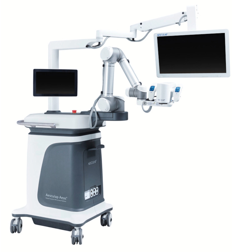
Objective: Current literature debates the role of newly developed three-dimensional (3D) Exoscopes in the daily routine of neurosurgical practice. So far, only a small number of cadaver lab studies or case reports have examined the novel Aesculap Aeos Three-Dimensional Robotic Digital Microscope. This study aims to evaluate the grade of satisfaction and intraoperative handling of this novel system in neurosurgery.

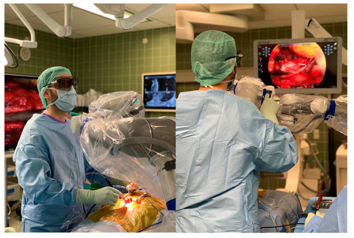
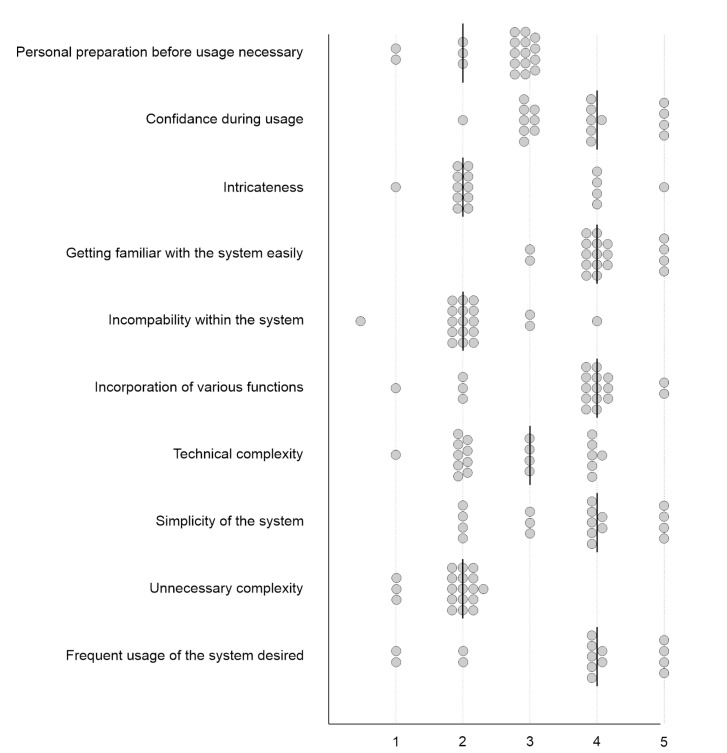
Methods: Nineteen neurosurgical procedures (12 cranial, 6 spinal and 1 peripheral nerve) performed over 9 weeks using the Aeos were analyzed. Ten neurosurgeons of varying levels of training were included after undergoing device instruction and training. Following every surgery, a questionnaire consisting of 43 items concerning intraoperative handling was completed. The questionnaires were analyzed using descriptive statistics.

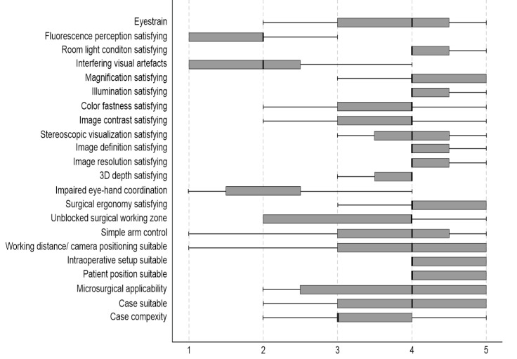
Results: No intraoperative complications occurred. Surgical satisfaction was ranked high (78.95%). In total, 84.21% evaluated surgical ergonomics as satisfactory, while 78.95% of the surgeons would like to use this system frequently. Image quality, independent working zoom function and depth of field were perceived as suboptimal by several neurosurgeons.

Conclusion: The use of Aeos is feasible and safe in microsurgical procedures, and surgical satisfaction was ranked high among most neurosurgeons in our study. The system might offer advanced ergonomic conditions in comparison to conventional ocular-based microscopes.

Keywords: Exoscope; ergonomics; microsurgery; operating microscope.


Conflict of interest statement

J.K. received honoraria from Aesculap AG, Brainlab AG, Carl Zeiss Meditec AG and CSL Behring GmbH. All other authors certify that they have no involvement in and no affiliations with any organization or entity with any financial interest (such as membership, educational grants, honoraria, consultancies, employment, stock ownership or other equity interests; patent-licensing or expert testimony agreements), or non-financial interests (such as professional or personal relationships, affiliations, knowledge or beliefs) concerning the subject matter or materials in this manuscript.

Figures

Figure 1
Figure 1
The surgeon during an operative resection of a meningioma TS 4/5 using the Three-Dimensional Robotic Digital Microscope.
Figure 2
Figure 2
The Aesculap Aeos Three-Dimensional Robotic Digital Microscope.
Figure 3
Figure 3
Screenshots of intraoperative recordings with Aeos during resection of a glioblastoma multiforme assisted by 5-ALA-induced PpIX fluorescence: (A) Conventional white light. The tumor and its margins are elusive under conventional white light. (B) Blue light. The tumor is clearly visible in strong fluorescence under dimmed white light and activated blue light. (C) Simultaneous usage of the white and blue light source: Tumor margins are easier to identify.
Figure 4
Figure 4
Intraoperative administration of indocyanine green during aneurysm clipping: Perception of indocyanine green during Aeos surgery in A and C. The unclipped aneurysm of the medial cerebral artery is dissected and visualized in (A). Visualization of the aneurysm under conventional light during surgery closed with a right-angled Sugita clip in (B). In (C) the light of the Aeos is on for post clipping control with indocyanine green. Rest perfusion in the clipped sack of the aneurysm at the right angle is highlighted with indocyanine green. Figure (DF) show even enhanced contrast by image processing, which however intraoperativly is not necessary due to high definition backlighted LED screen.
Figure 5
Figure 5
Evacuation of an intracerebral hemorrhage with the assistance of tubes. Intraoperative visualization of the hemorrhage during Aeos surgery.
Figure 6
Figure 6
Resection of an intraspinal, lumbar hemangioblastoma with Aeos. Visualization of the arachnoidea and spinal cord after dura opening during surgery in (A). Exposure of the underlying intradural tumor in (B).
Figure 7
Figure 7
A screenshot of the intraoperative recordings of vagus nerve stimulation in a patient suffering from therapy-resistant depression. The vagus nerve is illustrated in (A) during electrode placement. (B) represents the final view after electrode placement.
Figure 8
Figure 8
The first questionnaire evaluated the surgeon’s satisfaction regarding the applicability of Aeos. Factors influencing the applicability are listed on the y-axis. X-axis shows the level of agreement, where 0 is full disagreement, and 5 is full agreement. Boxplots illustrate the median agreement.
Figure 9
Figure 9
The second questionnaire evaluated the usability of Aeos. The attending surgeon rated the factors listed on the y-axis on a scale from 0, and 5 shown on the x-axis, where 0 is full disagreement, and 5 is full agreement. Dots illustrate ratings and lines illustrate medians.
Figure 10
Figure 10
The third questionnaire evaluated the strain of the performing surgeon. The surgeon rated the factors listed on the y-axis on a level of agreement between 0 and 20, illustrated on the x-axis, where 0 is full disagreement and 20 is full agreement. Boxplots illustrate median agreement.

References

    1. Amoo M., Henry J., Javadpour M. Beyond magnification and illumination: Preliminary clinical experience with the 4K 3D ORBEYE™ exoscope and a literature review. Acta Neurochir. 2021;163:2107–2115. doi: 10.1007/s00701-021-04838-8. - DOI - PubMed
    1. Khalessi A.A., Rahme R., Rennert R.C., Borgas P., Steinberg J.A., White T.G., Santiago-Dieppa D.R., Boockvar J.A., Hatefi D., Pannell J.S. First-in-man clinical experience using a high-definition 3-dimensional exoscope system for microneurosurgery. Oper. Neurosurg. 2019;16:717–725. doi: 10.1093/ons/opy320. - DOI - PubMed
    1. Langer D.J., White T.G., Schulder M., Boockvar J.A., Labib M., Lawton M.T. Advances in intraoperative optics: A brief review of current exoscope platforms. Oper. Neurosurg. 2020;19:84–93. doi: 10.1093/ons/opz276. - DOI - PubMed
    1. Murai Y., Sato S., Yui K., Morimoto D., Ozeki T., Yamaguchi M., Tateyama K., Nozaki T., Tahara S., Yamaguchi F. Preliminary clinical microneurosurgical experience with the 4K3-dimensional microvideoscope (ORBEYE) system for microneurological surgery: Observation study. Oper. Neurosurg. 2019;16:707–716. doi: 10.1093/ons/opy277. - DOI - PubMed
    1. Rösler J., Georgiev S., Roethe A.L., Chakkalakal D., Acker G., Dengler N.F., Prinz V., Hecht N., Faust K., Schneider U. Clinical implementation of a 3D4K-exoscope (Orbeye) in microneurosurgery. Neurosurg. Rev. 2021:1–9. doi: 10.1007/s10143-021-01577-3. - DOI - PMC - PubMed

LinkOut - more resources