. 2021 Aug 31;13(17):4395.
doi: 10.3390/cancers13174395.

Kallikrein 5 Inhibition by the Lympho-Epithelial Kazal-Type Related Inhibitor Hinders Matriptase-Dependent Carcinogenesis

Affiliations

Kallikrein 5 Inhibition by the Lympho-Epithelial Kazal-Type Related Inhibitor Hinders Matriptase-Dependent Carcinogenesis

Elaine Zayas Marcelino da Silva et al. Cancers (Basel). .

Abstract

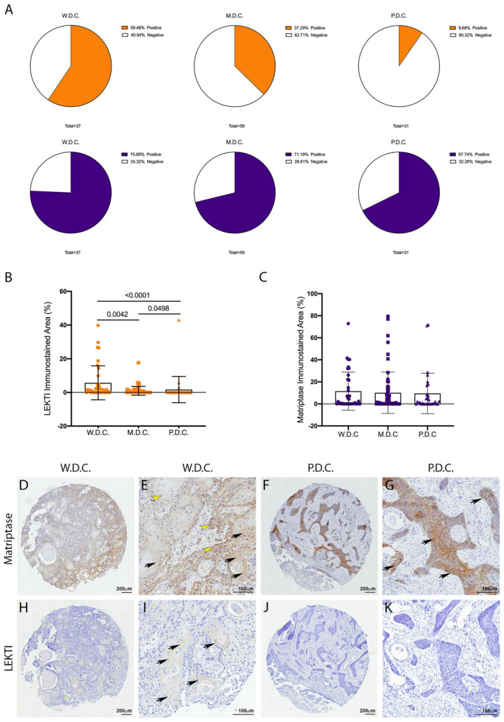
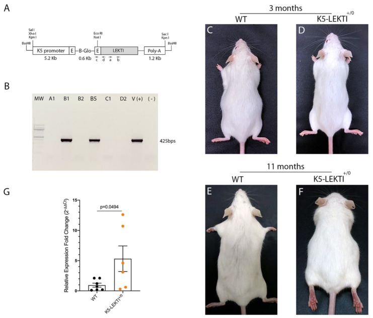
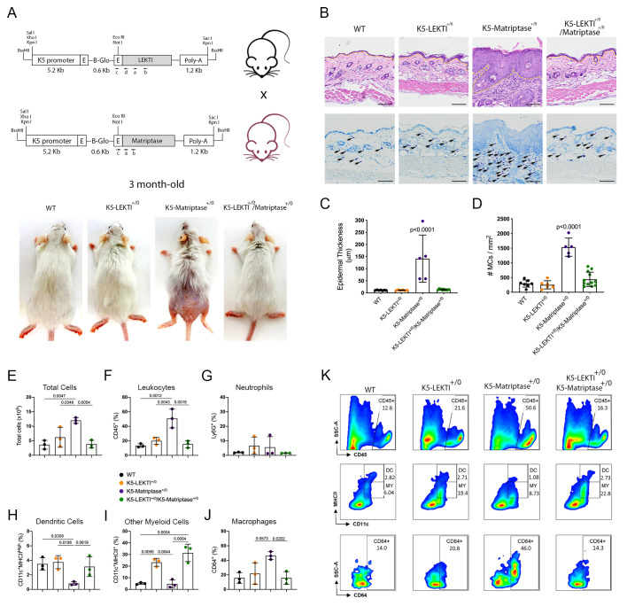
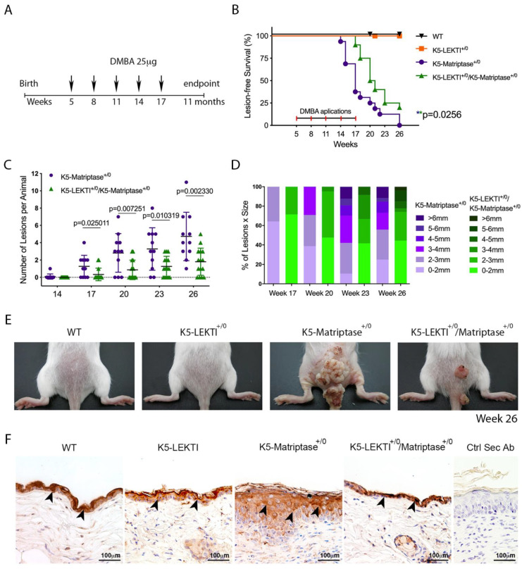
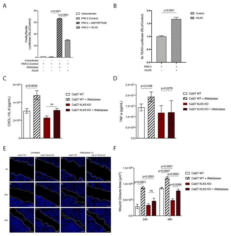
Head and neck squamous cell carcinoma remains challenging to treat with no improvement in survival rates over the past 50 years. Thus, there is an urgent need to discover more reliable therapeutic targets and biomarkers for HNSCC. Matriptase, a type-II transmembrane serine protease, induces malignant transformation in epithelial stem cells through proteolytic activation of pro-HGF and PAR-2, triggering PI3K-AKT-mTOR and NFKB signaling. The serine protease inhibitor lympho-epithelial Kazal-type-related inhibitor (LEKTI) inhibits the matriptase-driven proteolytic pathway, directly blocking kallikreins in epithelial differentiation. Hence, we hypothesized LEKTI could inhibit matriptase-dependent squamous cell carcinogenesis, thus implicating kallikreins in this process. Double-transgenic mice with simultaneous expression of matriptase and LEKTI under the keratin-5 promoter showed a prominent rescue of K5-Matriptase+/0 premalignant phenotype. Notably, in DMBA-induced SCC, heterotopic co-expression of LEKTI and matriptase delayed matriptase-driven tumor incidence and progression. Co-expression of LEKTI reverted altered Kallikrein-5 expression observed in the skin of K5-Matriptase+/0 mice, indicating that matriptase-dependent proteolytic pathway inhibition by LEKTI occurs through kallikreins. Moreover, we showed that Kallikrein-5 is necessary for PAR-2-mediated IL-8 release, YAP1-TAZ/TEAD activation, and matriptase-mediated oral squamous cell carcinoma migration. Collectively, our data identify a third signaling pathway for matriptase-dependent carcinogenesis in vivo. These findings are critical for the identification of more reliable biomarkers and effective therapeutic targets in Head and Neck cancer.

Keywords: KLK5; LEKTI; OSCC; SPINK5; matriptase.


Conflict of interest statement

The authors declare no potential conflict of interest.

Figures

Figure 1
Figure 1
Unlike LEKTI, matriptase is not modulated in poorly differentiated carcinomas. (A) IHC staining for LEKTI (orange) and matriptase (purple) in human OSCCs TMAs (W.D.C. n = 37, M.D.C. n = 59, and P.D.C. n = 31) showed that the number of positive samples for LEKTI (top panel) prominently decreases in the less differentiate samples, while for matriptase (bottom panel) this number remains similar. (B) Quantification of stained area confirmed that LEKTI is significantly decreased in M.D.Cs. and P.D.Cs.; p-values (One-Way ANOVA) are displayed in the graph. (C) Quantification of stained area shows that matriptase expression does not vary among W.D.Cs., M.D.Cs., and P.D.Cs.; (B,C) Data are expressed in mean ± SD. (DG) Representative images of matriptase IHC staining in well-differentiated and poorly differentiated carcinomas. (HK) Representative images of LEKTI IHC staining in well-differentiated and poorly differentiated carcinomas. Black arrows show deeper staining, while yellow arrowheads show diffuse staining. Lower magnifications (D,F,H,J)-bar = 100 μm; Higher magnifications (E,G,I,K) bar = 200 μm; Counterstaining with hematoxylin to visualize tissue architecture.
Figure 2
Figure 2
Generation of Keratin5-LEKTI transgenic mice. (A) Schematic structure of the K5-LEKTI transgene comprised of a bovine keratin-5 promoter (K5), rabbit-globin exons, a rabbit-globin intron, the mouse LEKTI cDNA and a rabbit-globin polyadenylation signal (PolyA). (a,b) position of primers used for qPCR and (c,d) genotyping. The linearized transgene vector was microinjected into the male pronucleus of FVB/NJ zygotes, which then were implanted into pseudopregnant mice. (B) LEKTI transgenic founders were genotyped by PCR using genomic DNA from tail biopsies with the primer pair indicated on the vector (c,d). Genotyping gel showing positive 425 bps amplified bands from founder mice. pBK5-LEKTI vector was used as template for positive control. (CF) Images of 3 (C,D) and 11 months (E,F) old WT (C,E) and K5-LEKTI+/0 (D,F) mice showing no differences on the outward phenotype. (G) qPCR analysis of the epidermis of newborn WT (black dots) and K5-LEKTI+/0 (orange dots) mice show a five-fold increase of LEKTI mRNA expression in transgenic compared to WT mice. WT n = 7 and K5-LEKTI+/0 n = 6; values are expressed in mean ± SD. p-values (two-tailed unpaired parametric t-test) are displayed in the graph.
Figure 3
Figure 3
Co-expression of LEKTI attenuates matriptase-mediated premalignant skin phenotype. (A) The scheme shows the breeding of K5-LEKTI+/0 with K5-Matriptase+/0 mice and the resulting litter of WT, K5-LEKTI+/0, K5-Matriptase+/0, and K5-Matriptase+/0/K5-LEKTI+/0 mice. Images show the outward appearance of these mice at 3 months of age. Matriptase-induced alopecia and ichthyosis are considerably attenuated by co-expression of LEKTI in Matriptase+/0/K5-LEKTI+/0. LEKTI+/0 in 3-month-old mice. (B) Representative histological appearance of dorsal skin of littermate WT (first column), K5-LEKTI+/0 (second column), K5-Matriptase+/0 (third column), and K5-Matriptase+/0/K5-LEKTI+/0 mice (forth column) stained by H&E (top panels) and Toluidine Blue (bottom panels) at 3 months of age. Bars = 100 μm. Yellow dashed lines show the limits between epidermis and dermis. Black arrows show metachromatically stained dermal mast cells. (C) Quantification of epidermal thickness in littermate WT (n = 7, black dots), K5-LEKTI+/0 (n = 6, orange dots), K5-Matriptase+/0 (n = 5, purple dots), and K5-Matriptase+/0/K5-LEKTI+/0 (n = 11, green dots) at 3 months of age. Data are expressed in mean ± SD. (D) Quantification of the dermal mast cell accumulation in the skin of littermate WT (black dots), K5-LEKTI+/0 (orange dots), K5-Matriptase+/0 (purple dots), and K5-Matriptase+/0/K5-LEKTI+/0 (green dots) at 3 months of age. Data are expressed in mean ± SD. (EJ) Myeloid cellular infiltration was evaluated in skin samples by flow cytometry. (E) Total cells (×105) were assessed by automated cell counter using trypan blue. The percentage of (F) leukocytes (CD45+), (G) neutrophils (Ly6G+ gated on CD45+), (H) dendritic cells (DC–CD11c+MHCIIHigh gated on CD45+Ly6G), (I) other myeloid cells (MY–CD11c+MHCII+ gated on CD45+Ly6G), and (J) macrophages (CD64+ gated on myeloid cells) in the skin. WT (black dots), K5-LEKTI+/0 (orange dots), K5-Matriptase+/0 (purple dots), and K5-Matriptase+/0/K5-LEKTI+/0 (green dots). (K) Representative flow cytometry of skin samples by groups. Data are representative of one experiment (n = 3/group) and are expressed as means ± SD. p-values (One-Way ANOVA with Tukey’s post-hoc test) displayed in the graphs.
Figure 4
Figure 4
Co-expression of LEKTI with matriptase in basal keratinocytes delays the onset and progression of chemically induced carcinogenesis. (A) One-stage chemical carcinogenesis scheme: dorsal skin of mice was exposed 5 times to 25 μg of DMBA, starting at week 5 of age, every 3 weeks, and were followed for up to 48 weeks of age. WT (n = 20), K5-LEKTI+/0 (n = 17), K5-Matriptase+/0 (n = 11), and K5-Matriptase+/0/K5-LEKTI+/0 (n = 16). (B) Kaplan–Meier analysis of tumor-free survival. WT (black upside-down triangle), K5-LEKTI+/0 (orange squares), K5-Matriptase+/0 (purple dots), and K5-Matriptase+/0/K5-LEKTI+/0 (green triangles). (C,D) Matriptase induced tumor progression in K5-Matriptase+/0, and K5-Matriptase+/0/K5-LEKTI+/0 mice. Data are expressed in mean ± SD. (C) number of lesions and (D) percentage of lesion of each size in littermate K5-Matriptase+/0 (purple dots) and K5-Matriptase+/0/K5-LEKTI+/0 (green triangles) mice from 14 to 26 weeks of age. p-values (multiple t-tests) are displayed in the graphs. (E) Representative images of the outward appearance of littermate WT, K5-LEKTI+/0, K5-Matriptase+/0, and K5-Matriptase+/0/K5-LEKTI+/0 mice at 26 weeks of age. (F) Klk5 IHC staining of the skin of 3-month-old WT, K5-LEKTI+/0, K5-Matriptase+/0, and K5-Matriptase+/0/LEKTI+/0 mice. Black arrowheads indicate stained areas; Negative secondary antibody control; Bar = 100 μm.
Figure 5
Figure 5
KLK5 activates YAP1-TAZ/TEAD transcription via PAR-2 and induces matriptase-mediated release of IL-8 and cell migration in OSCC cells. (A) KLK5-mediated PAR-2 activation analysis. HEK293T cells were transfected with pCDNA 3.1-PAR-2, pRL-Renilla luciferase and SRE-Firefly luciferase reporter plasmids and treated with hrKLK5 for 6 h, PAR-2 activation was measured by Luciferase activity. hrMatriptase was used as positive control for PAR-2 activation. p-values (One-Way ANOVA with Tukey’s post-hoc test) are displayed in the graphs. (B) Transcriptional activity of TEAD was also measured by luciferase assay with a reporter containing tandem TEAD-binding sites in HEK293T cells transfected with pCDNA 3.1-PAR-2 plasmid and treated with hrKLK5 for 6 h. p-values (two-tailed unpaired parametric t-test) are displayed in the graphs. (C,D) WT and KLK5 KO OSCC Cal 27 cell lines were serum-starved for 2 h, treated or not with hrMatriptase for 24 h, and proinflammatory cytokine release was evaluated by ELISA. (C) hrMatriptase treatment stimulated the release of CXCL-1/IL-8 in WT cells but not in KLK5 KO cells. (D) hrMatriptase treatment stimulated the release of TNF-α in both WT and KLK5 KO cells, although in KO cells this effect was very discreet. p-values (One-Way ANOVA with Tukey’s post-hoc test) are displayed in the graphs. (E,F) WT and KLK5 KO OSCC Cal 27 monolayers were serum-starved for 16 h, treated or not with hrMatriptase, scratched, and wound closure was evaluated for up to 48 h. (E) Representative images showing that hrMatriptase treatment induces wound closure in both WT and KLK5 KO cells (third and fourth columns) and this effect is partially inhibited in KLK5 KO cells (forth column) compared to WT cells (third column). Wound closure was also delayed in untreated KLK5 KO cells (second column) compared to WT cells (first column). (F) Quantification of the wound closure shows a delay in both matriptase treated (striped columns) and untreated (unstriped columns) KLK5 KO cells (red columns) when compared to WT cells (white columns) after 24 and 48 h. p-values (One-Way ANOVA with Tukey’s post-hoc test) are displayed in the graphs. (AF) Data are representative of two independent experiments and values are expressed in mean ± SEM.

References

    1. Bray F., Ferlay J., Soerjomataram I., Siegel R.L., Torre L.A., Jemal A. Global cancer statistics 2018: GLOBOCAN estimates of incidence and mortality worldwide for 36 cancers in 185 countries. CA Cancer J. Clin. 2018;68:394–424. doi: 10.3322/caac.21492. - DOI - PubMed
    1. Jemal A., Siegel R., Ward E., Hao Y., Xu J., Murray T., Thun M.J. Cancer statistics, 2008. CA Cancer J. Clin. 2008;58:71–96. doi: 10.3322/CA.2007.0010. - DOI - PubMed
    1. Molinolo A.A., Amornphimoltham P., Squarize C.H., Castilho R.M., Patel V., Gutkind J.S. Dysregulated molecular networks in head and neck carcinogenesis. Oral Oncol. 2009;45:324–334. doi: 10.1016/j.oraloncology.2008.07.011. - DOI - PMC - PubMed
    1. Forastiere A., Koch W., Trotti A., Sidransky D. Head and neck cancer. N. Engl. J. Med. 2001;345:1890–1900. doi: 10.1056/NEJMra001375. - DOI - PubMed
    1. Bagan J.V., Scully C. Recent advances in Oral Oncology 2007: Epidemiology, aetiopathogenesis, diagnosis and prognostication. Oral Oncol. 2008;44:103–108. doi: 10.1016/j.oraloncology.2008.01.008. - DOI - PubMed