

Peripapillary vessel density measurement of quadrant and clock-hour sectors in primary angle closure glaucoma using optical coherence tomography angiography
- PMID: 34503457
- PMCID: PMC8428096
- DOI: 10.1186/s12886-021-02093-0
Peripapillary vessel density measurement of quadrant and clock-hour sectors in primary angle closure glaucoma using optical coherence tomography angiography
Abstract
Background: The purpose of this study was to investigate diagnostic ability of peripapillary vessel density of primary angle closure glaucoma (PACG) eyes in quadrant and clock-hour sectors by optical coherence tomography angiography (OCTA).
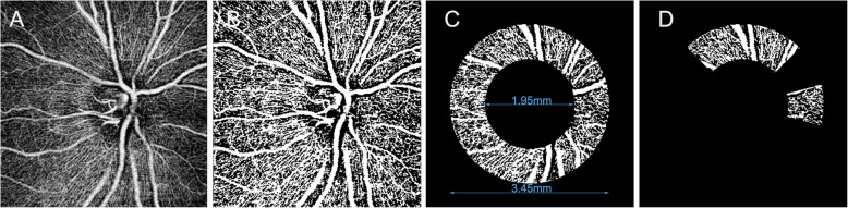
Methods: This was a cross-sectional study on forty-one PACG patients (41eyes) and twenty-seven healthy subjects (27 eyes). All subjects underwent OCTA (DRI OCT Triton; Topcon Corporation, Tokyo, Japan) and peripapillary retinal nerve fiber layer (RNFL) thickness imaging with swept-source optical coherence tomography (OCT). The peripapillary vessel density of quadrant and clock-hour sectors was quantified by imageJ software. The diagnostic capability of OCTA and OCT parameters was evaluated by the areas under the receiver operating characteristics curves (AUCs). Pearson correlation analysis or Spearman correlation test was used to evaluate the correlation between vessel density parameters and related factors.
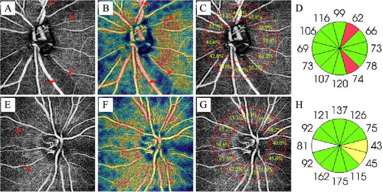
Results: Compared with the control group, the peripapillary vessel density of glaucomatous group was lower to different degrees in the four quadrants and each clock-hour sectors, and vessel density reduced most at 7 o'clock. The difference between the diagnostic ability of peripapillary vessel density and peripapillary RNFL thickness was not statistically significant, except 4 o'clock and inferior quadrant. The inferior quadrant peripapillary vessel density had the best diagnostic value (AUC0.969), followed by the 7 o'clock vessel density (AUC0.964), average vessel density (AUC0.939) and the 7 o'clock RNFL thickness (AUC0.919). The average peripapillary vessel density was correlated with average RNFL and visual field (VF) mean deviation (P < 0.001).
Conclusions: In PACG, the diagnostic ability of the peripapillary vessel density is equivalent to the peripapillary RNFL thickness. Understanding spatial characteristics of the peripapillary vessel density in PACG may be helpful for clinical diagnosis and monitoring the progress of diseases.
Keywords: Optical coherence tomography angiography; Peripapillary vessel density; Primary angle closure glaucoma.
© 2021. The Author(s).
Conflict of interest statement
The authors declare that they have no competing interests.
Figures
Similar articles
-
Diagnostic ability of peripapillary vessel density measurements of optical coherence tomography angiography in primary open-angle and angle-closure glaucoma.Br J Ophthalmol. 2017 Aug;101(8):1066-1070. doi: 10.1136/bjophthalmol-2016-309377. Epub 2016 Nov 29. Br J Ophthalmol. 2017. PMID: 27899368
-
Spatial positional relationship between macular superficial vessel density and ganglion cell-inner plexiform layer thickness in primary angle closure glaucoma.Int Ophthalmol. 2022 Jan;42(1):103-112. doi: 10.1007/s10792-021-02005-7. Epub 2021 Aug 15. Int Ophthalmol. 2022. PMID: 34392472 Free PMC article.
-
Changes of Optic Disc and Macular Vessel Perfusion Density in Primary Angle Closure Glaucoma: A Quantitative Study Using Optical Coherence Tomography Angiograph.Ophthalmic Res. 2023;66(1):1245-1253. doi: 10.1159/000533874. Epub 2023 Aug 30. Ophthalmic Res. 2023. PMID: 37647877 Free PMC article.
-
Optical coherence tomography angiography in glaucoma.Ann Transl Med. 2020 Sep;8(18):1204. doi: 10.21037/atm-20-2828. Ann Transl Med. 2020. PMID: 33241053 Free PMC article. Review.
-
Differentiating Degenerative from Vascular Dementia with the Help of Optical Coherence Tomography Angiography Biomarkers.Healthcare (Basel). 2022 Mar 15;10(3):539. doi: 10.3390/healthcare10030539. Healthcare (Basel). 2022. PMID: 35327019 Free PMC article. Review.
Cited by
-
Comparative of OCT and OCTA parameters in patients with early chronic angle-closure glaucoma and early pituitary adenoma.Sci Rep. 2024 Sep 13;14(1):21448. doi: 10.1038/s41598-024-71103-0. Sci Rep. 2024. PMID: 39271729 Free PMC article.
-
Vascular resistance indices are higher in the superior than inferior optic nerve head and retina.Exp Eye Res. 2024 Nov;248:110070. doi: 10.1016/j.exer.2024.110070. Epub 2024 Sep 5. Exp Eye Res. 2024. PMID: 39243927
-
The Topographic Relationship Between Choroidal Microvascular Dropout and Glaucomatous Damage in Primary Angle-Closure Glaucoma.Transl Vis Sci Technol. 2022 Oct 3;11(10):20. doi: 10.1167/tvst.11.10.20. Transl Vis Sci Technol. 2022. PMID: 36239967 Free PMC article.
-
Impact of acquisition area on deep-learning-based glaucoma detection in different plexuses in OCTA.Sci Rep. 2024 Sep 2;14(1):20414. doi: 10.1038/s41598-024-71235-3. Sci Rep. 2024. PMID: 39223266 Free PMC article.
-
Quantitative Measurements of Vessel Density and Blood Flow Areas Primary Angle Closure Diseases: A Study of Optical Coherence Tomography Angiography.J Clin Med. 2022 Jul 13;11(14):4040. doi: 10.3390/jcm11144040. J Clin Med. 2022. PMID: 35887804 Free PMC article.
References
-
- Rabiolo A, Gelormini F, Sacconi R, Cicinelli MV, Triolo G, Bettin P, Nouri-Mahdavi K, Bandello F, Querques G. Comparison of methods to quantify macular and peripapillary vessel density in optical coherence tomography angiography. PloS one. 2018;13(10):e0205773. doi: 10.1371/journal.pone.0205773. - DOI - PMC - PubMed
-
- Richter GM, Chang R, Situ B, Chu Z, Burkemper B, Reznik A, Bedrood S, Kashani AH, Varma R, Wang RK. Diagnostic performance of macular versus peripapillary vessel parameters by optical coherence tomography angiography for glaucoma. Transl Vis Sci Tech. 2018;7(6):21. doi: 10.1167/tvst.7.6.21. - DOI - PMC - PubMed
MeSH terms
Grants and funding
- 18-025/Joint Shantou International Eye Center of Shantou University and The Chinese University of Hong Kong
- 18-025/Joint Shantou International Eye Center of Shantou University and The Chinese University of Hong Kong
- 18-025/Joint Shantou International Eye Center of Shantou University and The Chinese University of Hong Kong
LinkOut - more resources
Full Text Sources
