Review
. 2021 Sep 14;78(11):1145-1165.
doi: 10.1016/j.jacc.2021.06.049.

Medial Arterial Calcification: JACC State-of-the-Art Review

Affiliations
Review

Medial Arterial Calcification: JACC State-of-the-Art Review

Peter Lanzer et al. J Am Coll Cardiol. .

Abstract

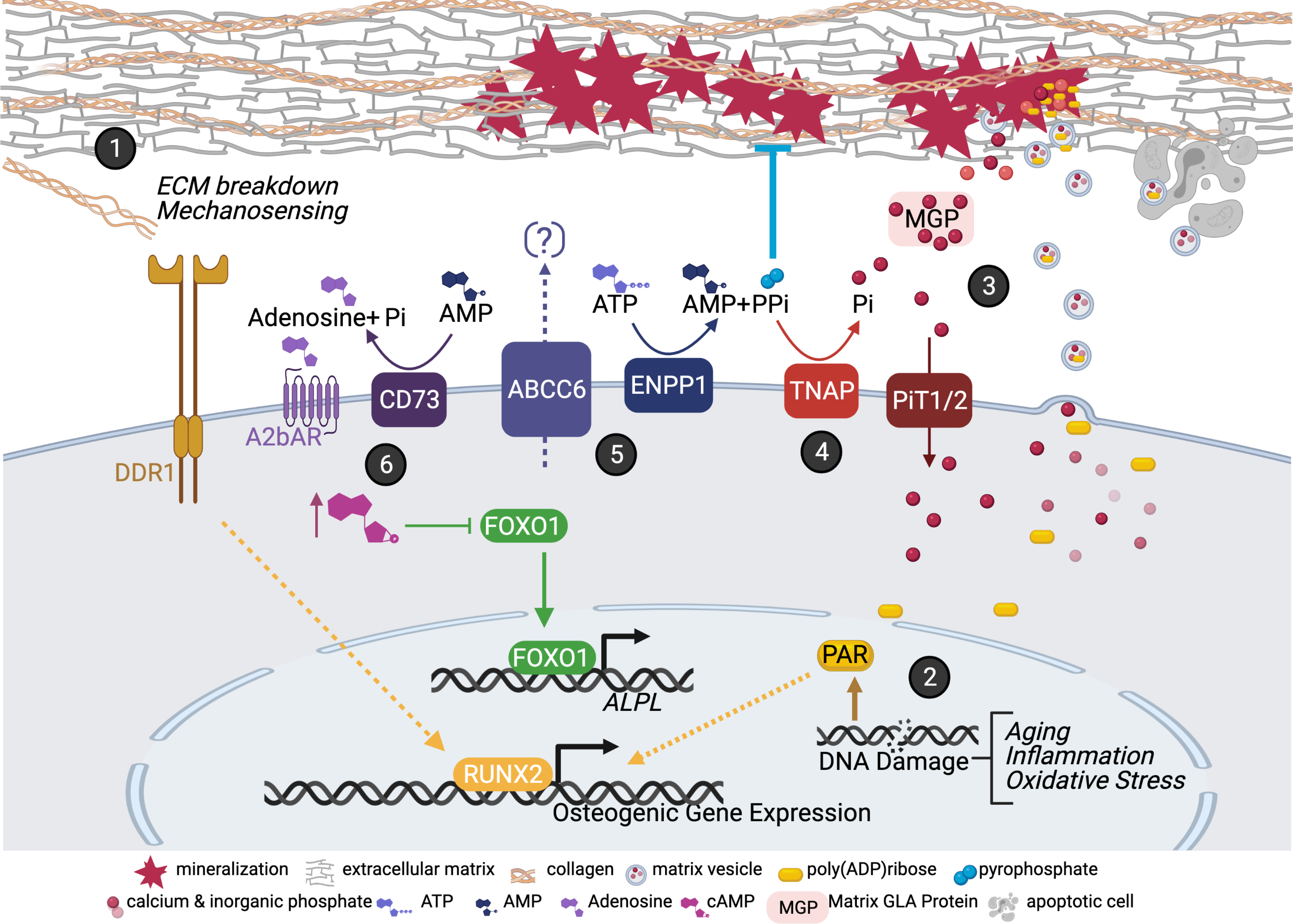
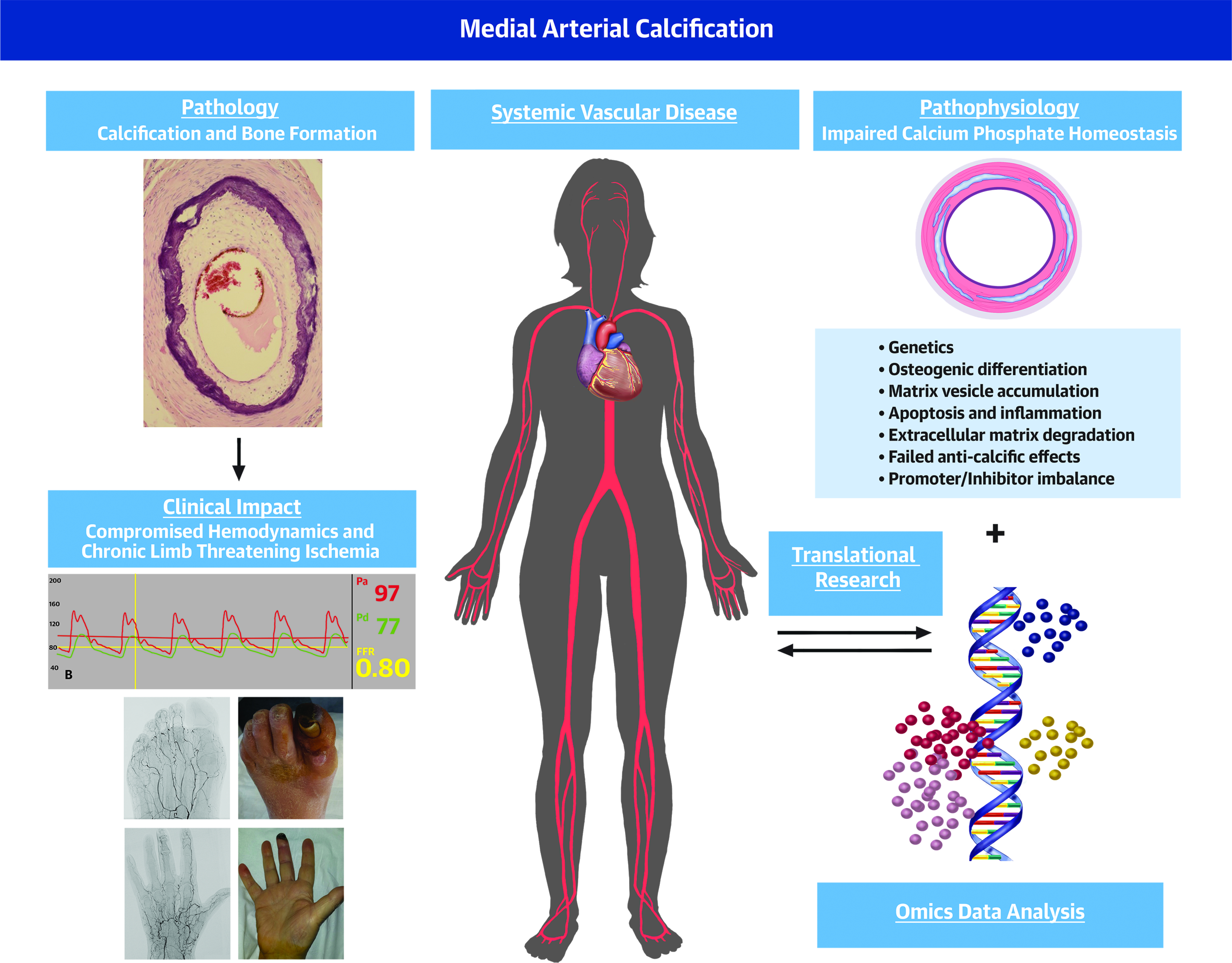
Medial arterial calcification (MAC) is a chronic systemic vascular disorder distinct from atherosclerosis that is frequently but not always associated with diabetes mellitus, chronic kidney disease, and aging. MAC is also a part of more complex phenotypes in numerous less common diseases. The hallmarks of MAC include disseminated and progressive precipitation of calcium phosphate within the medial layer, a prolonged and clinically silent course, and compromise of hemodynamics associated with chronic limb-threatening ischemia. MAC increases the risk of complications during vascular interventions and mitigates their outcomes. With the exception of rare monogenetic defects affecting adenosine triphosphate metabolism, MAC pathogenesis remains unknown, and causal therapy is not available. Implementation of genetics and omics-based approaches in research recognizing the critical importance of calcium phosphate thermodynamics holds promise to unravel MAC molecular pathogenesis and to provide guidance for therapy. The current state of knowledge concerning MAC is reviewed, and future perspectives are outlined.

Keywords: atherosclerosis; chronic limb-threatening ischemia; genetics; medial arterial calcification; omics; peripheral artery disease; vascular calcification.


Conflict of interest statement

Funding Support and Author Disclosures Dr Furniss is supported by the National Institute for Health Research (NIHR) Oxford Biomedical Research Centre. Dr Schuchardt has received support from Charité 3R and the Bundesministerium für Bildung und Forschung. Dr J.D. Lanzer has received support from Informatics for Life funded by the Klaus Tschira Foundation. Dr Thakker has received support from a Wellcome Trust Senior Investigator Award, an NIHR Senior Investigator Award, and the NIHR Oxford Biomedical Research Centre Programme. Dr Saez-Rodriguez has received support from Informatics for Life funded by the Klaus Tschira Foundation; has received funding from GSK and Sanofi; and expects consultant fees from Travere Therapeutics. Dr Millan has received financial support from the Spanish Ministry of Science, Innovation, and Universities (grant no: PGC2018_095795_B_I00). Dr Sato has received institutional research support from NIH-HL141425, Leducq Foundation Grant, 480 Biomedical, 4C Medical, 4Tech, Abbott, Accumedical, Alivas, Amgen, Biosensors, Boston Scientific, Canon USA, Cardiac Implants, Celonova, Claret Medical, Concept Medical, Cook, CSI, DuNing, Inc, Edwards LifeSciences, Emboline, Endotronix, Envision Scientific, Lutonix/Bard, Gateway, Lifetech, Limflo, MedAlliance, Medtronic, Mercator, Merill, Microport Medical, Microvention, Mitraalign, Mitra assist, NAMSA, Nanova, Neovasc, NIPRO, Novogate, Occulotech, OrbusNeich Medical, Phenox, Profusa, Protembis, Qool, ReCor Medical, Senseonics, Shockwave, Sinomed, Spectranetics, Surmodics, Symic, Vesper, W.L. Gore, and Xeltis. Dr Virmani has received institutional research support from NIH-HL141425, Leducq Foundation Grant, 480 Biomedical, 4C Medical, 4Tech, Abbott, Accumedical, Alivas, Amgen, Biosensors, Boston Scientific, Canon USA, Cardiac Implants, Celonova, Claret Medical, Concept Medical, Cook, CSI, DuNing, Inc, Edwards LifeSciences, Emboline, Endotronix, Envision Scientific, Lutonix/Bard, Gateway, Lifetech, Limflo, MedAlliance, Medtronic, Mercator, Merill, Microport Medical, Microvention, Mitraalign, Mitra assist, NAMSA, Nanova, Neovasc, NIPRO, Novogate, Occulotech, OrbusNeich Medical, Phenox, Profusa, Protembis, Qool, ReCor Medical, Senseonics, Shockwave, Sinomed, Spectranetics, Surmodics, Symic, Vesper, W.L. Gore, and Xeltis; is a consultant for Abbott Vascular, Boston Scientific, Celonova, OrbusNeich Medical, Terumo Corporation, W.L. Gore, Edwards Lifesciences, Cook Medical, CSI, ReCor Medical, SinoMedical Sciences Technology, Surmodics, and Bard BD; and is a Scientific Advisory Board Member for Medtronic and Xeltis. Dr St. Hilaire has received support from National Institutes of Health grants HL142932 and HL117917. All other authors have reported that they have no relationships relevant to the contents of this paper to disclose.

Figures

Figure 1.
Figure 1.. Mechanisms Contributing to MAC Pathogenesis.
(1) Extracellular matrix (ECM) cues are detected via DDR1 receptor promoting the activation of osteogenic genes. (2) DNA damage response pathway leads to the accumulation of poly (AMP) ribose (PAR) molecules. PAR are secreted in matrix vesicles (MVs) and act as niduses for calcification. (3) Calcium/phosphate minerals can nucleate in the ECM, but this is inhibited via MGLA. PiT1/2 transporter removes inorganic phosphate (Pi) from the extracellular environment. (4) TNAP converts the endogenous inhibitor of mineral nucleation, pyrophosphate (PPi), into the mineral building block, Pi. (5) PPi and AMP are produced from the activity of ENPP1 in the extracellular ATP metabolic pathway. The substrate for ABCC6 is thought to also be ATP but currently not known. (6) CD73-mediated A2b adenosine receptor signaling functions to suppress MAC via cAMP-mediated repression of FOXO1 gene. With a lack of this signaling FOXO1 upregulates ALPL/TNAP. Created
Figure 2.
Figure 2.. Histology of medial (MAC) and intimal (atherosclerosis) calcifications.
MAC and the intimal atherosclerotic calcifications are shown. The legends to each medial and intimal calcification pattern have been summarized in table 1. The histologic sections shown in the left and right columns of the medial and intimal calcifications were stained with Movat pentachrome (MP) (A-F and M-R) and hematoxylin and eosin (H&E) (G-L and S-X), respectively. The red boxes in the MP sections indicate areas of magnifications shown in the H&E sections and in stage IV (medial) and sheet (intimal) MP sections. IEL – internal elastic lamina (black arrows, black and white arrowheads). Modified and reproduced with permission from doi: 10.1016/j.jcmg.2018.08.039 and doi: 10.1016/j.ejvs.2020.08.037.
Figure 3.
Figure 3.. MAC and hemodynamics.
Male with diabetes mellitus and gangrene of the right middle finger and X-ray angiography demonstrating advanced MAC without evidence of stenotic lesions of the right upper extremity arteries. (A) High fidelity blood pressure recordings at the level of the mid brachial (Pa) and distal ulnar (Pd) arteries demonstrated Pd/Pa-ratio of 0.91 at baseline, (B) Pd/Pa-ratio of 0.80 following intravenous (140 μg/kg/min systemic infusion) and intra-arterial (150 μg) bolus administration of adenosine and (C) Pd/Pa-ratio of 0.76 following intra-arterial nitroglycerin (300 μg). The disproportional decrease in the distal diastolic pressures following vasodilatation appears to suggest marked microvascular dysfunction. Hemodynamically significant lesion proximal to the target territory has been excluded by X-ray angiography.
Figure 4.
Figure 4.. MAC in ultrasound.
Female with chronic kidney disease and MAC demonstrated by ultrasound. (A) B-mode ultrasound longitudinal image of the left superficial femoral artery (SFA) revealed on the near end arterial wall linear homogeneous echogenic band pathognomonic for MAC; note the smooth endothelial interface of the SFA; white arrowheads denote diffuse medial calcifications.(B) Color coded Doppler (CCD) image of A demonstrates normal arterial flow signal (blue color); red color corresponds to the accompanying vein; white arrowheads as in A. (C) CCD and pulse wave Doppler (PWD) images show a normal triphasic flow pattern indicating a normal artery.
Figure 5.
Figure 5.. MAC in native X-ray and X-ray angiographic images.
(A, B) Male with diabetes mellitus and MAC demonstrated by X-ray imaging. (A) native X-ray image of the right brachial artery demonstrating the typical “rail-road track” pattern of calcification parallel to the humerus, white arrowheads at the edge of calcifications (B) X-ray angiographic image of A showing the smooth endothelial interface following contrast media filling, heavy calcification are recognized as white stripes along the lumen (white arrowhead). (C, D) Male with cardiac arrhythmias undergoing coronary angiography. (C) High resolution native X-ray image of the right coronary artery reveals disseminated pattern of calcifications (two white arrowheads on the left) suggestive of MAC seen in A; the image appears partly duplicated due the rapid motion of the cardiac base (white arrowhead on the right). (D) X-ray coronary angiographic image of C showing the smooth endothelial interface demarcated by homogeneous contrast media filling (black arrow).
Figure 6.
Figure 6.. MAC in Computed Tomography.
3D CT reconstruction surface rendering in a male with MAC. (A) Whole body scan reveals calcifications (white spots) within the abdominal aorta, pelvic and leg arteries. (B) Enlarged section of the internal carotid artery from A appears relatively free from calcifications. (C) Enlarged section of the superficial femoral artery with heavy diffuse calcification typical for MAC. (D) Calcium windowing highlights the calcifications of the vasculature. (E) Coronary CT- angiography shows extensive calcifications of the proximal left anterior descending (LAD) coronary artery and lesser calcifications of the proximal right and left circumflex coronary arteries. (F) Enlarged section of the proximal LAD shows heavy confluent, partly circumferential calcifications.
Figure 7.
Figure 7.. MAC in Optical Coherence Tomography.
On OCT MAC appears as regions with homogeneous low signal intensity and sharply demarcated border zones. (A-C) Cross-sections of the coronary arteries. (A) MAC spans more than a half of the circumference (12 to 19 o’clock); (B) Extensive, partly eccentric MAC (13 to 15 o’clock); (C) Highly eccentric MAC (10 to 12 o’clock). Note the smooth and regular intima in all three images. Courtesy and Copyright, Abbott, Inc.
Figure 8.
Figure 8.. MAC in limb arteries.
Male with the gangrene of the great toe left. (A) Native X-ray image of the left foot; heavy calcifications of the metatarsal and toe arteries, particularly of the first metatarsal and great toe arteries are shown (white arrowheads). (B) Angiographic X-ray image corresponding to A; shown is the extensive destruction of the small arteries; paucity of the distal arterial supply is highlighted in the context of the great toe (oval shaped broken line). (C) Photograph of the left foot corresponding to A; shown is extensive necrosis of the left great toe. Male with the gangrene of the mid-finger left. (D) Native X-ray image of the left hand; heavy calcifications of the metacarpal and finger arteries are shown (white arrowheads). (E) Angiographic X-ray image corresponding to A; shown are multiple occlusions of the metacarpal and finger arteries and virtual absence of arterial blood flow to the fingers (exemplified by the mid-finger, oval shaped broken line). (F) Photograph of the left hand corresponding to A; shown is distal necrosis of the mid-finger left.
Figure 9.
Figure 9.. Calcified plaques and endovascular interventions (histology).
Examples of histological findings of intimal and medial calcifications following endovascular interventions. Upper panels: common femoral artery and superficial femoral artery (SFA) following drug eluting stent placement and orbital atherectomy device in ex vivo setting, respectively. Lower panels: coronary artery treated by rotational atherectomy device, SFA treated with intravascular lithotripsy device and plaque debris obtained from directional atherectomy device. Images A to D, H, I, L, M are stained with hematoxylin and eosin (H&E). Images E to G are stained with Movat pentachrome. Ca – calcifications; CTO – chronic total occlusion. Modified and reproduced with permission from DOI: 10.1016/j.jcin.2019.10.060. Images F and G: modified and reproduced with permission from doi: 10.1016/0002-8703(95)90384-4.
Figure 10.
Figure 10.. MAC and Omics.
Overview of pathophysiology of MAC and the proposed Omics approaches to study MAC. (Left box) Different proposed pathophysiologic principles suggested triggering and promoting the vascular calcifications including MAC. (Middle section) Calcified sites in the vasculature can be studied with omics at different degrees of resolutions. Bulk omics (bottom) provide a gross tissue profiles. Single cell omics (middle) provide profiles of individual cells. Spatial omics (top) yields profiles of regions represented as the 2D sections of the tissues. (Right box) The proposed approaches have not been systematically applied to study MAC but hold promise to address numerous aspects of calcifications processes providing tangible insights and hypotheses for molecular pathogenesis of MAC.
Central Illustration.
Central Illustration.. MAC: A Systemic Vascular Disorder Devastating Peripheral Circulation.
. In health calcium phosphate (CaP) homeostasis is maintained and crystallization prevented. In MAC CaP homeostasis brakes down resulting in progressive mineralization and in more advanced stages, bone formation within the medial layer. A number of pathogenetic principles including smooth muscle cells osteogenic differentiation, apoptosis, inflammation, molecular defects of matrisome have been reported to regulate the calcification process (left panels). MAC impairs hemodynamics often causing chronic limb threatening ischemia (right panels). Omics approaches holds distinct promise to define MAC molecular pathogenesis and design treatments (central panels).

References

    1. Reesink KD, Spronck B. Constitutive interpretation of arterial stiffness in clinical studies: A methodological review. Am J Physiol Heart Circ Physiol 2019;316:H693–H709. - PubMed
    1. Weber T, Chirinos JA. Pulsatile arterial haemodynamics in heart failure. Eur Heart J 2018;39:3847–3854. - PMC - PubMed
    1. Chirinos JA, Segers P, Hughes T, Townsend R. Large-artery stiffness in health and Disease; JACC State-of-the-Art review. J Am Coll Cardiol 2019;74:1237–1263. - PMC - PubMed
    1. Mowafy KA, Soliman M, Hammoda AM, Soliman RM. Bilateral lower limb disabling claudication in a young man: A case of Mönckeberg’s arteriosclerosis. Vasc Endovasc Rev https://www.verjournal.com/articles/Disabling-Claudication-Monckebergs-A... (June 6, 2020).
    1. Pisani I, De Troia A, Allegri L, Corradi D, Vaglio A. Malignant Mönckeberg medial calcific sclerosis. Intern Emerg Med 2018;13:615–617. - PubMed

Publication types