. 2021 Oct;42(10):1904-1911.
doi: 10.3174/ajnr.A7250. Epub 2021 Sep 9.

A Longitudinal Analysis of Early Lesion Growth in Presymptomatic Patients with Cerebral Adrenoleukodystrophy

Affiliations

A Longitudinal Analysis of Early Lesion Growth in Presymptomatic Patients with Cerebral Adrenoleukodystrophy

E J Mallack et al. AJNR Am J Neuroradiol. 2021 Oct.

Abstract

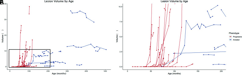
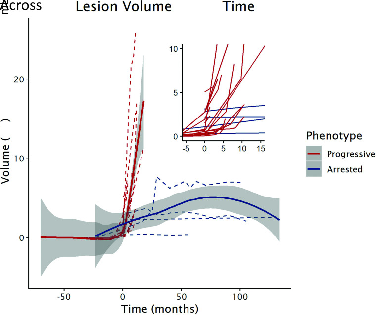
Background and purpose: Cerebral adrenoleukodystrophy is a devastating neurological disorder caused by mutations in the ABCD1 gene. Our aim was to model and compare the growth of early cerebral lesions from longitudinal MRIs obtained in presymptomatic patients with progressive and arrested cerebral adrenoleukodystrophy using quantitative MR imaging-based lesion volumetry.

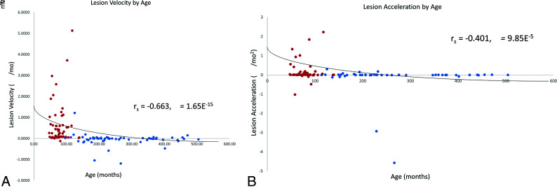
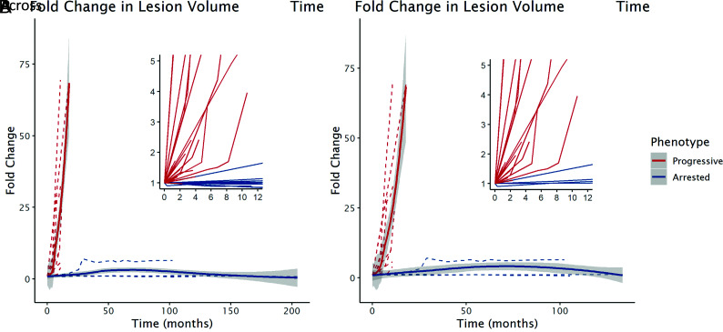
Materials and methods: We retrospectively quantified and modeled the longitudinal growth of early cerebral lesions from 174 MRIs obtained from 36 presymptomatic male patients with cerebral adrenoleukodystrophy. Lesions were manually segmented using subject-specific lesion-intensity thresholding. Volumes were calculated and plotted across time. Lesion velocity and acceleration were calculated between sequentially paired and triplet MRIs, respectively. Linear mixed-effects models were used to assess differences in growth parameters between progressive and arrested phenotypes.

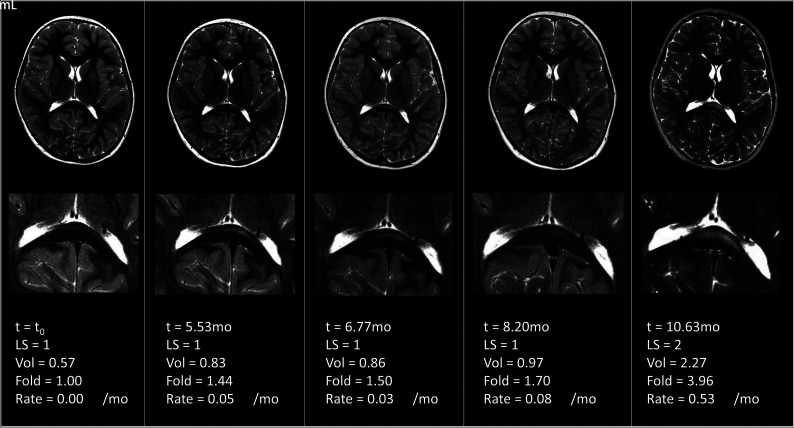
Results: The median patient age was 7.4 years (range, 3.9-37.0 years). Early-stage cerebral disease progression was inversely correlated with age (ρ = -0.6631, P < .001), early lesions can grow while appearing radiographically stable, lesions undergo sustained acceleration in progressive cerebral adrenoleukodystrophy (β = 0.10 mL/month2 [95% CI, 0.05-0.14 mL/month2], P < .001), and growth trajectories diverge between phenotypes in the presymptomatic time period.

Conclusions: Measuring the volumetric changes in newly developing cerebral lesions across time can distinguish cerebral adrenoleukodystrophy phenotypes before symptom onset. When factored into the overall clinical presentation of a patient with a new brain lesion, quantitative MR imaging-based lesion volumetry may aid in the accurate prediction of patients eligible for therapy.


Figures

FIG 1.
FIG 1.
A, Patient-specific lesion trajectories of each cohort plotted against patient age. B, Zoomed view of outlined area in 1A): 0–18 years of age on the x-axis and 0- to 10-mL lesion volume on the y-axis. Note the exponential growth trajectories for patients with progressive CALD.
FIG 2.
FIG 2.
A, Lesion growth velocities over sequential MR imaging pairs and B, acceleration over sequential MR imaging triplets, stratified by age in patients with progressive CALD (red) and arrested CALD (blue). Presymptomatic lesion progression is inversely correlated with age (P < .001).
FIG 3.
FIG 3.
Serial MRIs demonstrating accelerated lesion growth without a change in the LS in patient 11.
FIG 4.
FIG 4.
A, Lesion growth trajectories from the time of the first abnormal MR imaging finding in presymptomatic patients with progressive (n = 23, red) versus arrested (n = 13, blue) CALD. Lesion volume increased 2.5× per month (fold change β = 2.49 [95% CI, 2.10–2.89], P < .001) in the patients with progressive CALD. B, In the cohort of patients with an initial LS ≤ 2 (restricted cohort; n = 19 progressive, n = 4 arrested), lesion growth was similar (β = 2.54 [95% CI, 2.04–3.04], P < .001) for the patients with progressive CALD.
FIG 5.
FIG 5.
Nonparametric local-weighted regression curves demonstrating the early lesion growth trajectories between phenotypes measured by raw lesion volume across time in the cohort of patients with an initial LS ≤ 2.

References

    1. Moser HW. Adrenoleukodystrophy: phenotype, genetics, pathogenesis and therapy. Brain 1997;120:1485–1508 10.1093/brain/120.8.1485 - DOI - PubMed
    1. Huffnagel IC, Laheji FK, Aziz-Bose R, et al. . The natural history of adrenal insufficiency in X-linked adrenoleukodystrophy: an international collaboration. J Clin Endocrinol Metab 2019;104:118–26 10.1210/jc.2018-01307 - DOI - PubMed
    1. Moser HW, Loes DJ, Melhem ER, et al. . X-linked adrenoleukodystrophy: overview and prognosis as a function of age and brain magnetic resonance imaging abnormality—a study involving 372 patients. Neuropediatrics 2000;31:227–39 10.1055/s-2000-9236 - DOI - PubMed
    1. Liberato AP, Mallack EJ, Aziz-Bose R, et al. . MRI brain lesions in asymptomatic boys with X-linked adrenoleukodystrophy. Neurology 2019;92:e1698–1708 10.1212/WNL.0000000000007294 - DOI - PMC - PubMed
    1. Loes DJ, Fatemi A, Melhem ER, et al. . Analysis of MRI patterns aids prediction of progression in X-linked adrenoleukodystrophy. Neurology 2003;61:369–74 10.1212/01.wnl.0000079050.91337.83 - DOI - PubMed

Publication types

LinkOut - more resources