

Structural analysis of receptors and actin polarity in platelet protrusions
- PMID: 34504018
- PMCID: PMC8449362
- DOI: 10.1073/pnas.2105004118
Structural analysis of receptors and actin polarity in platelet protrusions
Abstract
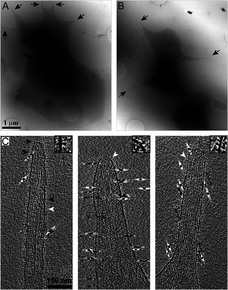
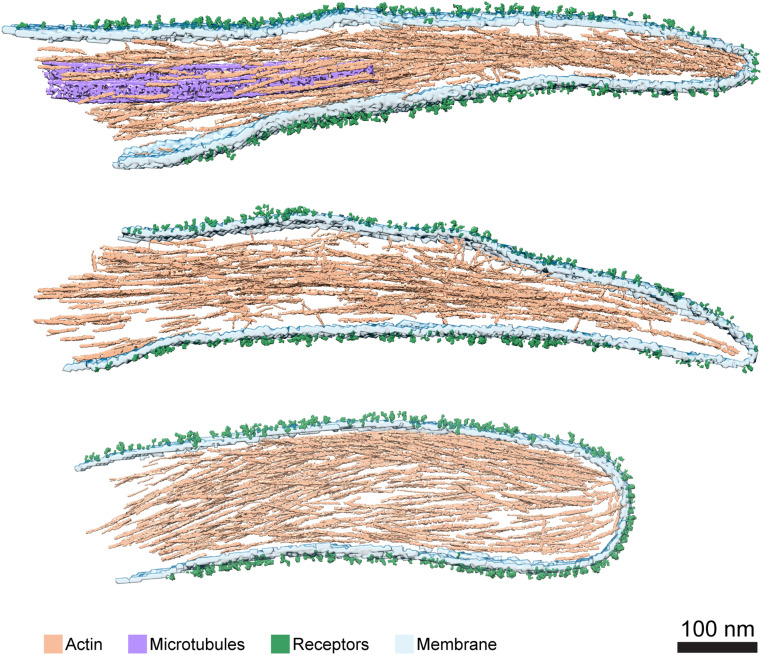
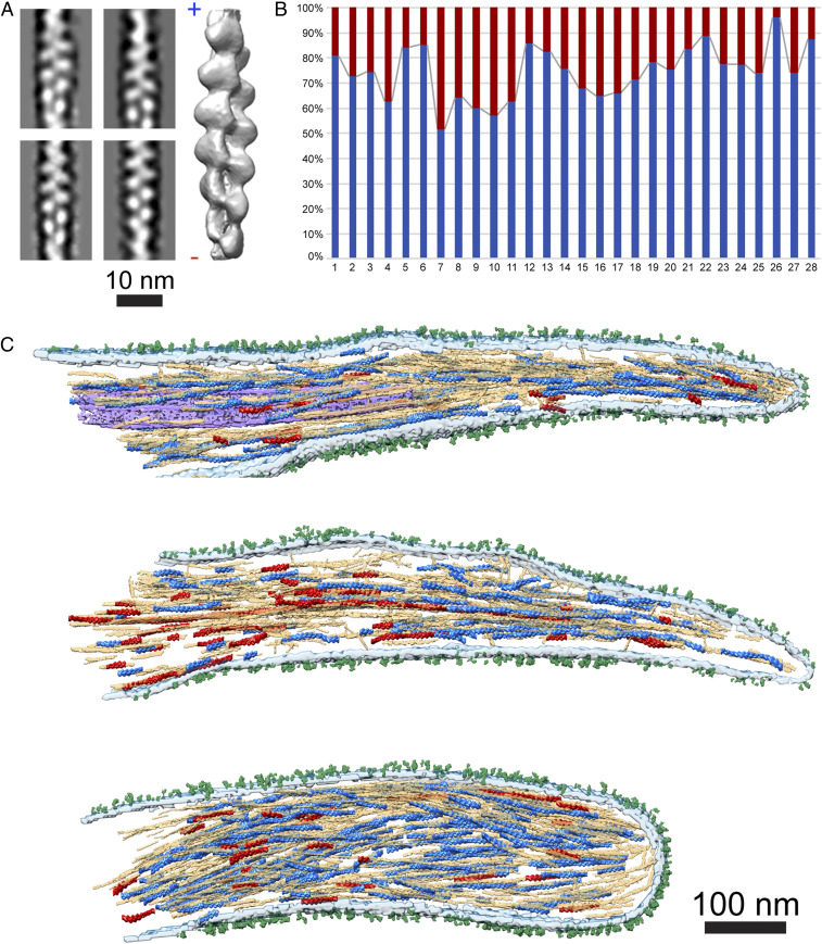
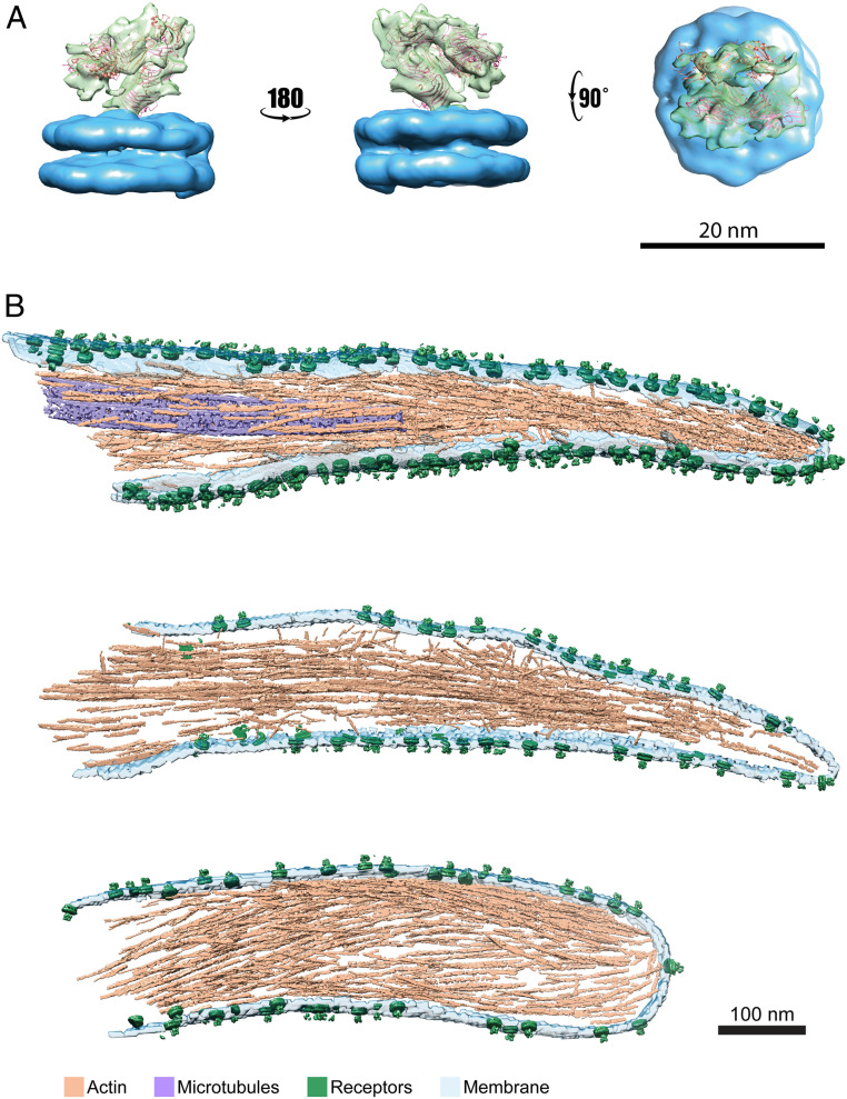
During activation the platelet cytoskeleton is reorganized, inducing adhesion to the extracellular matrix and cell spreading. These processes are critical for wound healing and clot formation. Initially, this task relies on the formation of strong cellular-extracellular matrix interactions, exposed in subendothelial lesions. Despite the medical relevance of these processes, there is a lack of high-resolution structural information on the platelet cytoskeleton controlling cell spreading and adhesion. Here, we present in situ structural analysis of membrane receptors and the underlying cytoskeleton in platelet protrusions by applying cryoelectron tomography to intact platelets. We utilized three-dimensional averaging procedures to study receptors at the plasma membrane. Analysis of substrate interaction-free receptors yielded one main structural class resolved to 26 Å, resembling the αIIbβ3 integrin folded conformation. Furthermore, structural analysis of the actin network in pseudopodia indicates a nonuniform polarity of filaments. This organization would allow generation of the contractile forces required for integrin-mediated cell adhesion.
Keywords: actin; cryoelectron tomography; platelets; receptors.
Copyright © 2021 the Author(s). Published by PNAS.
Conflict of interest statement
The authors declare no competing interest.
Figures
Similar articles
-
Calpain regulation of cytoskeletal signaling complexes in von Willebrand factor-stimulated platelets. Distinct roles for glycoprotein Ib-V-IX and glycoprotein IIb-IIIa (integrin alphaIIbbeta3) in von Willebrand factor-induced signal transduction.J Biol Chem. 1997 Aug 29;272(35):21847-54. doi: 10.1074/jbc.272.35.21847. J Biol Chem. 1997. PMID: 9268316
-
Role of G protein signaling in the formation of the fibrin(ogen)-integrin αIIbβ3-actin cytoskeleton complex in platelets.Platelets. 2016 Sep;27(6):563-75. doi: 10.3109/09537104.2016.1147544. Epub 2016 Mar 30. Platelets. 2016. PMID: 27026498
-
Synergistic effect of signaling from receptors of soluble platelet agonists and outside-in signaling in formation of a stable fibrinogen-integrin αIIbβ3-actin cytoskeleton complex.Thromb Res. 2015 Jan;135(1):114-20. doi: 10.1016/j.thromres.2014.10.005. Epub 2014 Oct 14. Thromb Res. 2015. PMID: 25456731
-
On the structure and function of platelet integrin alpha IIb beta 3, the fibrinogen receptor.Proc Soc Exp Biol Med. 1995 Apr;208(4):346-60. doi: 10.3181/00379727-208-43863a. Proc Soc Exp Biol Med. 1995. PMID: 7535429 Review.
-
Platelet adhesion receptors.Semin Cell Biol. 1995 Oct;6(5):305-14. doi: 10.1006/scel.1995.0040. Semin Cell Biol. 1995. PMID: 8562923 Review.
Cited by
-
Platelets and diseases: signal transduction and advances in targeted therapy.Signal Transduct Target Ther. 2025 May 16;10(1):159. doi: 10.1038/s41392-025-02198-8. Signal Transduct Target Ther. 2025. PMID: 40374650 Free PMC article. Review.
-
Characterizing Integrin Tensions during Platelet Adhesion and Stiffness Sensing.Nano Lett. 2025 Jul 16;25(28):11156-11163. doi: 10.1021/acs.nanolett.5c02578. Epub 2025 Jul 3. Nano Lett. 2025. PMID: 40605589
-
A network of mixed actin polarity in the leading edge of spreading cells.Commun Biol. 2022 Dec 7;5(1):1338. doi: 10.1038/s42003-022-04288-7. Commun Biol. 2022. PMID: 36473943 Free PMC article.
-
Organization, dynamics and mechanoregulation of integrin-mediated cell-ECM adhesions.Nat Rev Mol Cell Biol. 2023 Feb;24(2):142-161. doi: 10.1038/s41580-022-00531-5. Epub 2022 Sep 27. Nat Rev Mol Cell Biol. 2023. PMID: 36168065 Free PMC article. Review.
-
The cellular protrusions for inter-cellular material transfer: similarities between filopodia, cytonemes, tunneling nanotubes, viruses, and extracellular vesicles.Front Cell Dev Biol. 2024 Jul 5;12:1422227. doi: 10.3389/fcell.2024.1422227. eCollection 2024. Front Cell Dev Biol. 2024. PMID: 39035026 Free PMC article. Review.
References
-
- Patel D., et al. ., Dynamics of GPIIb/IIIa-mediated platelet-platelet interactions in platelet adhesion/thrombus formation on collagen in vitro as revealed by videomicroscopy. Blood 101, 929–936 (2003). - PubMed
-
- Jackson S. P., Nesbitt W. S., Westein E., Dynamics of platelet thrombus formation. J. Thromb. Haemost. 7 (suppl. 1), 17–20 (2009). - PubMed
-
- Lebowitz E. A., Cooke R., Contractile properties of actomyosin from human blood platelets. J. Biol. Chem. 253, 5443–5447 (1978). - PubMed
Publication types
MeSH terms
Substances
LinkOut - more resources
Full Text Sources
Other Literature Sources
