. 2021 Sep 9;11(1):17918.
doi: 10.1038/s41598-021-95958-9.

Prognostic role of Ki-67 in glioblastomas excluding contribution from non-neoplastic cells

Affiliations

Prognostic role of Ki-67 in glioblastomas excluding contribution from non-neoplastic cells

Rikke H Dahlrot et al. Sci Rep. .

Abstract

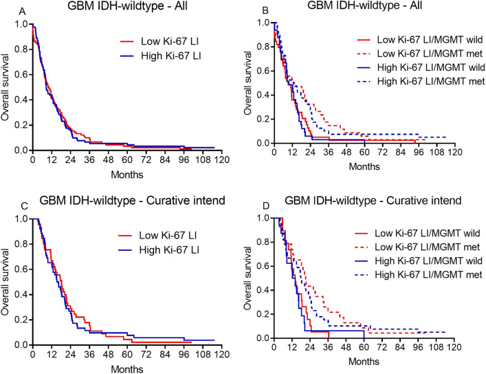
Survival of glioblastoma patients varies and prognostic markers are important in the clinical setting. With digital pathology and improved immunohistochemical multiplexing becoming a part of daily diagnostics, we investigated the prognostic value of the Ki-67 labelling index (LI) in glioblastomas more precisely than previously by excluding proliferation in non-tumor cells from the analysis. We investigated the Ki-67 LI in a well-annotated population-based glioblastoma patient cohort (178 IDH-wildtype, 3 IDH-mutated). Ki-67 was identified in full tumor sections with automated digital image analysis and the contribution from non-tumor cells was excluded using quantitative double-immunohistochemistry. For comparison of the Ki-67 LI between WHO grades (II-IV), 9 IDH-mutated diffuse astrocytomas and 9 IDH-mutated anaplastic astrocytomas were stained. Median Ki-67 LI increased with increasing WHO grade (median 2.7%, 6.4% and 27.5%). There was no difference in median Ki-67 LI between IDH-mutated and IDH-wildtype glioblastomas (p = 0.9) and Ki-67 LI was not associated with survival in glioblastomas in neither univariate (p = 0.9) nor multivariate analysis including MGMT promoter methylation status and excluding IDH-mutated glioblastomas (p = 0.2). Ki-67 may be of value in the differential diagnostic setting, but it must not be over-interpreted in the clinico-pathological context.


Conflict of interest statement

The authors declare no competing interests.

Figures

Figure 1
Figure 1
Immunohistochemical expression of Ki-67 (brown) and the exclusion-markers CD45, CD31, Iba1 and ASMA (red)—identified by a double immunohistochemical staining cocktail in astrocytic brain tumors. (A) IDH-mutated diffuse astrocytoma (DA) with low cellularity showing only a few scattered Ki-67 positive tumor cells not labeled by red (see insert). (B) IDH-mutated anaplastic astrocytoma (AA) with moderate cellularity showing higher presence of Ki-67 positive tumor cells not labeled by red. (C) IDH-mutated glioblastoma (GBM) and (D) IDH-wildtype glioblastoma with high cellularity and increased number of Ki-67 positive tumor cells not labeled by red. An increased fraction of non-tumor cells also expressed Ki-67 (see insert in C). The appearance of pseudopalisading necrosis with pink positive staining of microglia/macrophages (black asterix) and microvascular proliferations with pink positive staining of endothelial/smooth muscle cells (black arrow) are shown respectively in (C) and (D). (E + F) The staining was quantified by a software-based classifier identifying tumor cells having Ki-67 positive nuclei and not being labeled by red (blue arrowhead, blue label), non-tumor cells having Ki-67 positive nuclei and red cytoplasm (turquoise arrowhead, gray label) and supposed Ki-67 negative tumor cells not being labeled by red (green label). Magnification by × 20 (A-D) and × 80 (EF). Scale bar indicates 100 µm (A-D), 25 µm (EF).
Figure 2
Figure 2
Box-plots showing the Ki-67 LI in different glioma subgroups. The horizontal line is the median.
Figure 3
Figure 3
Kaplain Meier curves shown for all patients (A) and patients with known MGMT promoter status (B). Similar curves are shown for patients receiving radiotherapy 59.4 Gy on 33 fraction and concomitant and adjuvant Temozolomide in (C) and (D).

References

    1. Zhou YH, et al. Establishment of prognostic models for astrocytic and oligodendroglial brain tumors with standardized quantification of marker gene expression and clinical variables. Biomark. Insights. 2010;5:153–168. doi: 10.4137/bmi.s6167. - DOI - PMC - PubMed
    1. Wrensch M, et al. Diagnostic, treatment, and demographic factors influencing survival in a population-based study of adult glioma patients in the San Francisco Bay Area. Neuro Oncol. 2006;8:12–26. doi: 10.1215/s1522851705000268. - DOI - PMC - PubMed
    1. Tsidulko AY, et al. Prognostic relevance of NG2/CSPG4, CD44 and Ki-67 in patients with glioblastoma. Tumour Boil. 2017;39:1010428317724282. doi: 10.1177/1010428317724282. - DOI - PubMed
    1. Hegi ME, et al. Correlation of O6-methylguanine methyltransferase (MGMT) promoter methylation with clinical outcomes in glioblastoma and clinical strategies to modulate MGMT activity. J. Clin. Oncol. 2008;26:4189–4199. doi: 10.1200/JCO.2007.11.5964. - DOI - PubMed
    1. Malmstrom A, et al. Temozolomide versus standard 6-week radiotherapy versus hypofractionated radiotherapy in patients older than 60 years with glioblastoma: The Nordic randomised, phase 3 trial. Lancet Oncol. 2012;13:916–926. doi: 10.1016/s1470-2045(12)70265-6. - DOI - PubMed

Publication types