

Diagnostic classification of coronavirus disease 2019 (COVID-19) and other pneumonias using radiomics features in CT chest images
- PMID: 34504246
- PMCID: PMC8429652
- DOI: 10.1038/s41598-021-97497-9
Diagnostic classification of coronavirus disease 2019 (COVID-19) and other pneumonias using radiomics features in CT chest images
Abstract
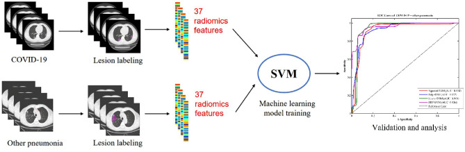
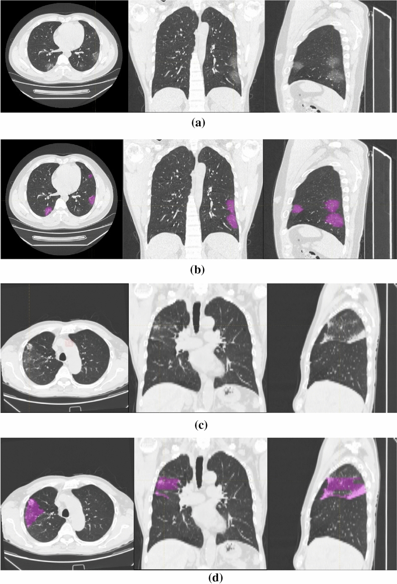
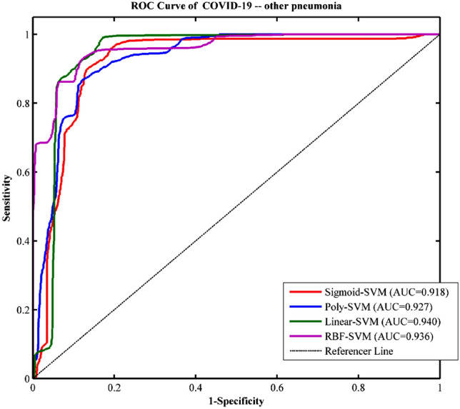
We propose a classification method using the radiomics features of CT chest images to identify patients with coronavirus disease 2019 (COVID-19) and other pneumonias. The chest CT images of two groups of participants (90 COVID-19 patients who were confirmed as positive by nucleic acid test of RT-PCR and 90 other pneumonias patients) were collected, and the two groups of data were manually drawn to outline the region of interest (ROI) of pneumonias. The radiomics method was used to extract textural features and histogram features of the ROI and obtain a radiomics features vector from each sample. Then, we divided the data into two independent radiomic cohorts for training (70 COVID-19 patients and 70 other pneumonias patients), and validation (20 COVID-19 patients and 20 other pneumonias patients) by using support vector machine (SVM). This model used 20 rounds of tenfold cross-validation for training. Finally, single-shot testing of the final model was performed on the independent validation cohort. In the COVID-19 patients, correlation analysis (multiple comparison correction-Bonferroni correction, P < 0.05/7) was also conducted to determine whether the textural and histogram features were correlated with the laboratory test index of blood, i.e., blood oxygen, white blood cell, lymphocytes, neutrophils, C-reactive protein, hypersensitive C-reactive protein, and erythrocyte sedimentation rate. The final model showed good discrimination on the independent validation cohort, with an accuracy of 89.83%, sensitivity of 94.22%, specificity of 85.44%, and AUC of 0.940. This proved that the radiomics features were highly distinguishable, and this SVM model can effectively identify and diagnose patients with COVID-19 and other pneumonias. The correlation analysis results showed that some textural features were positively correlated with WBC, and NE, and also negatively related to SPO2H and NE. Our results showed that radiomic features can classify COVID-19 patients and other pneumonias patients. The SVM model can achieve an excellent diagnosis of COVID-19.
© 2021. The Author(s).
Conflict of interest statement
The authors declare no competing interests.
Figures
Similar articles
-
Machine learning-based CT radiomics model distinguishes COVID-19 from non-COVID-19 pneumonia.BMC Infect Dis. 2021 Sep 8;21(1):931. doi: 10.1186/s12879-021-06614-6. BMC Infect Dis. 2021. PMID: 34496794 Free PMC article.
-
A COVID-19 risk score combining chest CT radiomics and clinical characteristics to differentiate COVID-19 pneumonia from other viral pneumonias.Aging (Albany NY). 2021 Mar 13;13(7):9186-9224. doi: 10.18632/aging.202735. Epub 2021 Mar 13. Aging (Albany NY). 2021. PMID: 33713401 Free PMC article.
-
A novel CT-based radiomics in the distinction of severity of coronavirus disease 2019 (COVID-19) pneumonia.BMC Infect Dis. 2021 Jun 25;21(1):608. doi: 10.1186/s12879-021-06331-0. BMC Infect Dis. 2021. PMID: 34171991 Free PMC article.
-
Thoracic imaging tests for the diagnosis of COVID-19.Cochrane Database Syst Rev. 2020 Nov 26;11:CD013639. doi: 10.1002/14651858.CD013639.pub3. Cochrane Database Syst Rev. 2020. Update in: Cochrane Database Syst Rev. 2021 Mar 16;3:CD013639. doi: 10.1002/14651858.CD013639.pub4. PMID: 33242342 Updated.
-
Artificial intelligence model on chest imaging to diagnose COVID-19 and other pneumonias: A systematic review and meta-analysis.Eur J Radiol Open. 2022;9:100438. doi: 10.1016/j.ejro.2022.100438. Epub 2022 Aug 18. Eur J Radiol Open. 2022. PMID: 35996746 Free PMC article. Review.
Cited by
-
Immunohematologic Biomarkers in COVID-19: Insights into Pathogenesis, Prognosis, and Prevention.Pathog Immun. 2023 Jun 26;8(1):17-50. doi: 10.20411/pai.v8i1.572. eCollection 2023. Pathog Immun. 2023. PMID: 37427016 Free PMC article. Review.
-
A Novel COVID-19 Diagnosis Approach Utilizing a Comprehensive Set of Diagnostic Information (CSDI).J Clin Med. 2023 Nov 3;12(21):6912. doi: 10.3390/jcm12216912. J Clin Med. 2023. PMID: 37959377 Free PMC article.
-
A review on advances in 18F-FDG PET/CT radiomics standardisation and application in lung disease management.Insights Imaging. 2022 Feb 5;13(1):22. doi: 10.1186/s13244-021-01153-9. Insights Imaging. 2022. PMID: 35124733 Free PMC article. Review.
-
Development and Validation of a Radiomics Nomogram Using Computed Tomography for Differentiating Immune Checkpoint Inhibitor-Related Pneumonitis From Radiation Pneumonitis for Patients With Non-Small Cell Lung Cancer.Front Immunol. 2022 Apr 26;13:870842. doi: 10.3389/fimmu.2022.870842. eCollection 2022. Front Immunol. 2022. PMID: 35558076 Free PMC article.
-
BND-VGG-19: A deep learning algorithm for COVID-19 identification utilizing X-ray images.Knowl Based Syst. 2022 Dec 22;258:110040. doi: 10.1016/j.knosys.2022.110040. Epub 2022 Oct 21. Knowl Based Syst. 2022. PMID: 36284666 Free PMC article.
References
MeSH terms
Substances
LinkOut - more resources
Full Text Sources
Medical
Research Materials
